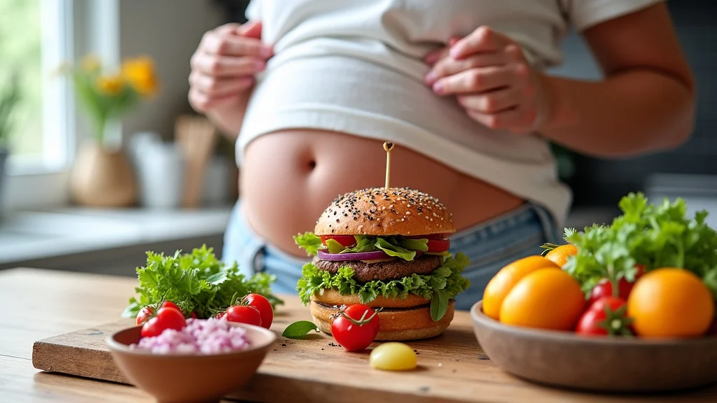
So, you’re hitting the second trimester and suddenly thinking about what to eat. It’s common to feel a little unsure about creating a good second trimester meal plan. You want to give your growing baby all the right nutrients, but you also might be dealing with new energy levels or cravings.
Don’t worry, it’s simpler than it seems! This guide will break it down step-by-step, making it easy for you to feel confident about your food choices.
Key Takeaways
- Understanding the nutritional needs of the second trimester is key for healthy growth.
- This meal plan focuses on balanced meals with essential vitamins and minerals.
- Simple meal ideas help combat common pregnancy discomforts and cravings.
- Hydration is just as important as food for your well-being.
- Snack ideas are included to keep energy levels stable throughout the day.
- This plan offers flexibility to fit your personal tastes and schedule.
Nourishing Your Second Trimester Body
The second trimester is often called the “golden period” of pregnancy. Many of the early pregnancy symptoms like morning sickness start to fade, and you might feel a surge of energy. This is a great time to focus on building a healthy eating pattern to support your baby’s rapid development and your own changing body.
Your baby is growing quickly, and they need plenty of good stuff from you. This means paying attention to protein, healthy fats, and essential vitamins and minerals.
Why a Second Trimester Meal Plan Matters
Creating a structured second trimester meal plan can help you stay on track with your nutrition. Without a plan, it’s easy to fall back on less healthy convenience foods, especially when you’re busy or feeling tired. A plan helps ensure you’re getting a wide variety of nutrients consistently.
It also helps manage cravings by having healthier options readily available. Think of it as a roadmap to feeling your best and providing the best for your baby.
Essential Nutrients for the Second Trimester
During this phase, your body needs more energy and specific nutrients to support your baby’s growth and your increased blood volume. Key among these are protein, which builds your baby’s tissues, and iron, crucial for carrying oxygen to your baby and preventing anemia. Calcium and Vitamin D are vital for bone development in your baby, and folate remains important for preventing neural tube defects.
Iron is a mineral that helps your body make hemoglobin, the protein in red blood cells that carries oxygen. Many women find their iron needs increase significantly in the second trimester. Good sources include lean red meat, poultry, fish, beans, and fortified cereals.
Pairing iron-rich foods with Vitamin C sources, like citrus fruits or bell peppers, can help your body absorb iron better.
Calcium is another vital nutrient for your baby’s developing bones and teeth. It also plays a role in your own bone health. Dairy products, leafy green vegetables like kale, and fortified plant-based milks are excellent sources.
Vitamin D works with calcium to help the body absorb it effectively. You can get Vitamin D from fatty fish, fortified milk, and by spending some time in the sun, though dietary sources are often more reliable.
Folate, also known as folic acid when supplemented, is incredibly important for preventing birth defects of the brain and spine. While it’s crucial throughout pregnancy, it remains a key nutrient in the second trimester. Leafy green vegetables, legumes, and fortified grains are good dietary sources.
Many healthcare providers recommend a daily prenatal vitamin that includes folic acid to ensure adequate intake.
- Protein helps build your baby’s muscles and tissues. Aim for about 70-100 grams per day.
- Iron is essential for blood production for both you and your baby. Your recommended intake often doubles during pregnancy.
- Calcium and Vitamin D are critical for your baby’s bone and teeth formation.
- Folate continues to be important for your baby’s brain and spinal cord development.
Crafting Your Second Trimester Meal Plan
Designing your second trimester meal plan doesn’t have to be complicated. The goal is to create balanced meals that include a good mix of protein, healthy fats, complex carbohydrates, and plenty of fruits and vegetables. This approach ensures you get a wide range of nutrients and helps keep your energy levels stable throughout the day, preventing those energy dips that can lead to unhealthy snacking.
Building Balanced Meals
A balanced meal typically includes a lean protein source, a whole grain or complex carbohydrate, and plenty of vegetables or fruits. For example, a lunch could be grilled chicken breast with a side of quinoa and a large mixed green salad. Dinner might be baked salmon with roasted sweet potatoes and steamed broccoli.
These combinations provide sustained energy and a variety of vitamins and minerals.
Protein sources can include chicken, turkey, fish (low-mercury options like salmon, trout, cod), lean beef, eggs, beans, lentils, tofu, and Greek yogurt. Whole grains offer fiber and sustained energy; think brown rice, oats, whole wheat bread, and quinoa. Healthy fats are crucial for brain development and hormone production; avocados, nuts, seeds, and olive oil are excellent choices.
Vegetables and fruits are packed with vitamins, minerals, and fiber. Aim for a rainbow of colors to ensure you’re getting a broad spectrum of nutrients. Dark leafy greens, berries, citrus fruits, and colorful vegetables like bell peppers and carrots are great additions to any meal or snack.
When planning, think about variety. Eating the same few meals every day can lead to nutrient gaps and boredom. Try to incorporate different types of proteins, grains, and produce throughout the week to maximize your nutrient intake and keep your meals interesting.
Sample Meal Plan Ideas
Here are some simple ideas to get you started. Remember, these are just suggestions, and you can adjust them based on your preferences and what’s available.
Breakfast Ideas
- Oatmeal with berries, nuts, and a drizzle of honey.
- Scrambled eggs with spinach and whole-wheat toast.
- Greek yogurt with granola and sliced fruit.
- Smoothie made with spinach, banana, protein powder, and almond milk.
Oatmeal provides fiber to keep you full and steady energy. Adding berries gives you antioxidants and Vitamin C, while nuts offer healthy fats and protein. Scrambled eggs are a fantastic source of protein and choline, important for your baby’s brain development.
Whole-wheat toast adds complex carbohydrates for energy. Greek yogurt is rich in protein and calcium. Smoothies are a quick way to pack in nutrients, especially if you’re not feeling very hungry.
Lunch Ideas
- Lentil soup with a side of whole-grain bread.
- Tuna salad (made with Greek yogurt instead of mayo) on whole-wheat crackers with carrot sticks.
- Chicken breast salad with mixed greens, cucumber, tomatoes, and a vinaigrette dressing.
- Quinoa bowl with black beans, corn, avocado, and salsa.
Lentil soup is packed with protein and fiber, making it very filling. Whole-grain bread offers complex carbs. Tuna salad provides protein and omega-3 fatty acids, though it’s best to consume it in moderation due to mercury concerns; opt for light tuna.
Using Greek yogurt instead of mayonnaise adds protein and reduces fat. A chicken breast salad offers lean protein and a wide array of vitamins from the vegetables. The quinoa bowl is a complete meal with protein, fiber, and healthy fats.
Dinner Ideas
- Baked salmon with roasted sweet potatoes and asparagus.
- Lean ground turkey stir-fry with brown rice and mixed vegetables.
- Chicken breast with a side of steamed broccoli and a small baked potato.
- Vegetarian chili with a dollop of Greek yogurt.
Salmon is a powerhouse of omega-3 fatty acids, crucial for your baby’s brain and eye development. Sweet potatoes are rich in Vitamin A and fiber. Turkey stir-fry is a lean protein option with plenty of opportunities to add various vegetables for nutrients and fiber.
Brown rice provides whole grains. Chicken breast is a versatile lean protein. Broccoli is a cruciferous vegetable packed with vitamins and fiber.
Vegetarian chili is a great way to get protein and fiber from beans and lentils.
Snack Smart
Snacks are essential for keeping your energy up and preventing you from getting overly hungry, which can lead to poor food choices. Aim for snacks that combine protein and fiber for sustained fullness. About 2-3 snacks per day are usually recommended.
- A handful of almonds and a piece of fruit.
- Apple slices with peanut butter.
- Hard-boiled eggs.
- Carrot sticks with hummus.
- A small container of cottage cheese or Greek yogurt.
Almonds offer healthy fats, protein, and fiber. Fruits provide natural sweetness and vitamins. Apple slices with peanut butter combine fiber, vitamins, and protein for a satisfying snack.
Hard-boiled eggs are a simple, portable source of protein. Carrot sticks with hummus provide fiber and plant-based protein. Cottage cheese and Greek yogurt are excellent sources of protein and calcium, helping to keep you satisfied between meals.
Staying Hydrated And Managing Cravings
Hydration is just as crucial as your diet during pregnancy. It helps prevent constipation, fatigue, and headaches, and it’s vital for amniotic fluid production and nutrient transport to your baby. Don’t forget that a good second trimester meal plan includes plenty of fluids!
The Importance of Water
Your body needs more fluids during pregnancy, especially in the second trimester. Aim for at least 8-10 glasses of water per day. If plain water feels boring, try infusing it with fruits like lemon, cucumber, or berries.
Herbal teas (check with your doctor about safe options) can also contribute to your fluid intake.
Dehydration can lead to uncomfortable symptoms like dizziness and fatigue. It can also increase your risk of urinary tract infections, which are more common during pregnancy. So, keep a water bottle with you throughout the day and sip on it regularly.
Listen to your body; if you feel thirsty, drink up!
Other healthy fluid options include milk (for calcium and protein), unsweetened juices in moderation, and clear broths. However, water should always be your primary beverage choice. Limit sugary drinks like soda and excessively caffeinated beverages, as they offer little nutritional value and can contribute to unwanted weight gain or interfere with sleep.
Handling Pregnancy Cravings
Cravings are a normal part of pregnancy. Your body might be signaling a need for certain nutrients, or it could just be hormonal. The key is to find a balance.
If you crave something sweet, try a piece of fruit or a small portion of dark chocolate instead of a large candy bar.
If you’re craving salty snacks, opt for air-popped popcorn, whole-grain crackers with a little cheese, or a small portion of mixed nuts. For crunchy cravings, try raw vegetables like carrots, celery, or bell peppers. Having healthier alternatives readily available makes it easier to satisfy your cravings without derailing your nutrition.
It’s okay to indulge occasionally in things you truly desire, but focus on mindful eating. Enjoy the treat in a smaller portion and savor it. If a craving is persistent, try to incorporate it into a balanced meal or snack.
For instance, if you’re craving ice cream, have a small scoop after a nutritious dinner. This way, you still get the satisfaction without overdoing it.
Tracking your cravings can sometimes reveal patterns. Are you craving certain foods when you’re tired or stressed? Addressing the underlying feeling might help reduce the intensity of the craving.
Sometimes, simply drinking a glass of water can help distinguish between thirst and a true craving.
Sample Week-Long Second Trimester Meal Plan
Here’s a sample week to give you a more concrete idea of how to structure your meals. This plan is rich in nutrients and designed for variety.
Monday
- Breakfast: Oatmeal with sliced banana and walnuts.
- Lunch: Large salad with grilled chicken, mixed greens, cucumber, bell peppers, and a light vinaigrette.
- Dinner: Baked cod with steamed broccoli and a side of brown rice.
- Snacks: Apple slices with peanut butter, a handful of almonds.
This day focuses on lean protein and whole grains. Cod is a good source of lean protein and omega-3s. Broccoli offers vitamins and fiber.
The snacks provide sustained energy and healthy fats.
Tuesday
- Breakfast: Scrambled eggs with spinach and whole-wheat toast.
- Lunch: Lentil soup with whole-grain bread.
- Dinner: Turkey meatballs with whole wheat pasta and marinara sauce, served with a side salad.
- Snacks: Greek yogurt with berries, carrot sticks with hummus.
Lentils are a fantastic source of protein and iron. Turkey meatballs provide lean protein, and whole wheat pasta offers complex carbohydrates. Greek yogurt is a good calcium and protein source.
Wednesday
- Breakfast: Smoothie with kale, banana, protein powder, and almond milk.
- Lunch: Quinoa bowl with black beans, corn, avocado, and salsa.
- Dinner: Grilled salmon with roasted sweet potatoes and green beans.
- Snacks: Hard-boiled egg, orange slices.
This day emphasizes omega-3s from salmon and complex carbohydrates from quinoa and sweet potatoes. Kale adds a dose of vitamins and minerals.
Thursday
- Breakfast: Greek yogurt with granola and mixed berries.
- Lunch: Tuna salad (made with Greek yogurt) on whole-grain crackers with cherry tomatoes.
- Dinner: Chicken breast stir-fry with mixed vegetables (broccoli, carrots, bell peppers) and brown rice.
- Snacks: Pear slices, small portion of mixed nuts.
Tuna provides protein and omega-3s. The stir-fry is a great way to load up on vegetables and lean protein. Mixed nuts are good for healthy fats and fiber.
Friday
- Breakfast: Whole wheat pancakes with a small amount of maple syrup and a side of scrambled eggs.
- Lunch: Leftover chicken stir-fry.
- Dinner: Homemade whole wheat pizza with lean protein (chicken or turkey sausage) and plenty of vegetables.
- Snacks: Cottage cheese with pineapple chunks, cucumber slices.
Pancakes can be a weekend treat, balanced with protein from eggs. Pizza can be a healthier option when made with whole wheat and lean toppings. Cottage cheese is rich in casein protein for slow release.
Saturday
- Breakfast: Oatmeal with peaches and chia seeds.
- Lunch: Veggie burger on a whole wheat bun with a side salad.
- Dinner: Lean beef stew with root vegetables (carrots, potatoes, parsnips).
- Snacks: A small handful of grapes, a hard-boiled egg.
Chia seeds add fiber and omega-3s. A veggie burger provides plant-based protein and fiber. Beef stew offers iron and protein, with nutrient-rich root vegetables.
Sunday
- Breakfast: Omelette with mushrooms, onions, and a slice of whole-wheat toast.
- Lunch: Leftover beef stew.
- Dinner: Roasted chicken with a baked potato and steamed mixed vegetables.
- Snacks: Banana with a tablespoon of almond butter, a small container of Greek yogurt.
This day offers a balanced mix of protein, complex carbs, and vitamins. Almond butter provides healthy fats and protein. A roasted chicken dinner is a classic, nutritious meal.
Common Myths Debunked
Myth 1: You Need To Eat For Two
While your calorie needs increase in the second trimester, it’s not double. You typically need an extra 300-350 calories per day, not an extra full person’s worth. Focusing on nutrient-dense foods is more important than simply increasing the quantity of food consumed.
Myth 2: All Seafood Is Bad Due To Mercury
This is not true. Many types of fish are safe and highly beneficial during pregnancy, offering essential omega-3 fatty acids. It’s important to choose low-mercury options like salmon, trout, sardines, and shrimp, while limiting high-mercury fish like shark, swordfish, and king mackerel.
Always check local advisories for fish caught in local waters.
Myth 3: You Can’t Eat Any Dairy
Dairy products are an excellent source of calcium and vitamin D, which are vital for your baby’s bone development. As long as the dairy products are pasteurized, they are safe and recommended. Unpasteurized dairy can carry harmful bacteria.
So, enjoy pasteurized milk, cheese, and yogurt!
Myth 4: Craving Specific Foods Means You’re Deficient In That Nutrient
While some cravings might hint at a need for certain nutrients, it’s often more complex. Hormonal changes, food aversions from morning sickness, and psychological factors can all play a role. It’s best to focus on a balanced diet overall rather than trying to satisfy every craving with specific foods, as this can lead to an unbalanced intake.
Frequently Asked Questions
Question: How much weight should I expect to gain in the second trimester?
Answer: Typically, a healthy weight gain during the second trimester is about one pound per week, or roughly 12-15 pounds over the entire trimester. However, this can vary, and it’s best to discuss your specific weight gain with your healthcare provider.
Question: Are there any specific vitamins I should focus on in the second trimester?
Answer: While a balanced diet covers many needs, iron, calcium, Vitamin D, and folate remain particularly important. Your doctor may recommend a prenatal vitamin to ensure you’re getting enough of these and other essential nutrients.
Question: What if I’m experiencing heartburn?
Answer: Heartburn is common in the second trimester. Eating smaller, more frequent meals, avoiding lying down immediately after eating, and avoiding trigger foods like spicy or greasy items can help. Your doctor can also suggest safe remedies.
Question: Can I eat spicy food during the second trimester?
Answer: For most healthy pregnancies, spicy foods are generally safe. However, they can sometimes trigger heartburn or indigestion, so listen to your body. If spicy food makes you uncomfortable, it’s best to limit it.
Question: How do I make sure I’m getting enough protein?
Answer: Include a protein source at every meal and snack. Good sources include lean meats, poultry, fish, eggs, dairy products, beans, lentils, tofu, and nuts. Aim for about 70-100 grams of protein daily.
Wrap Up
Creating a healthy second trimester meal plan helps you nourish yourself and your baby. Focus on balanced meals with lean protein, whole grains, healthy fats, and plenty of fruits and vegetables. Stay hydrated and manage cravings with smart choices.
You’ve got this!