Author: Admin

  • Second Trimester Is When Key Changes Happen

    Second Trimester Is When Key Changes Happen

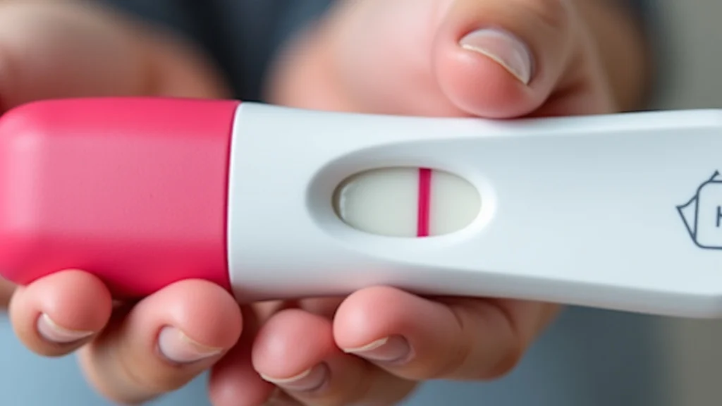
    Many people starting out find the phrase “second trimester is when” a bit confusing. It sounds like a big deal, and it is, but it doesn’t have to be scary. This period in pregnancy brings lots of new feelings and developments.

    We will walk through it step by step. You’ll learn what to expect and feel more prepared for this exciting time. Get ready to explore what makes the second trimester so special.

    Key Takeaways

    • You will learn what the second trimester officially is.
    • Discover the common signs and symptoms experienced during this time.
    • Understand the important medical check-ups and tests scheduled.
    • Explore tips for staying healthy and comfortable during these months.
    • Learn about the baby’s significant growth and development milestones.
    • Understand how to prepare for the upcoming changes and the final trimester.

    What Is the Second Trimester

    The second trimester is a very distinct phase of pregnancy. It generally starts around week 13 and goes until the end of week 28. Many pregnant people find this period more comfortable than the first.

    The intense morning sickness often fades, and energy levels tend to rise. This is when your baby really starts to grow and develop quickly. It’s a time filled with noticeable changes for both you and your little one.

    Beginning of the Second Trimester

    The transition from the first trimester to the second often feels like a welcome relief. The fatigue and nausea that can plague early pregnancy usually start to lessen. You might begin to feel more like yourself again.

    This renewed energy can be a great boost.

    • Energy levels typically increase after the first trimester fatigue.
    • Morning sickness often subsides or becomes much less severe.
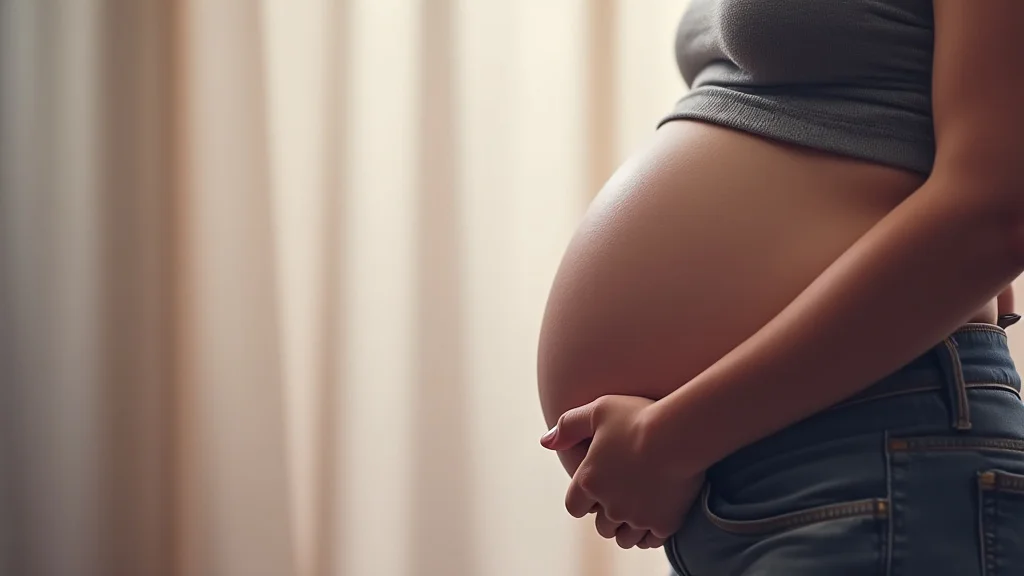
    • Some women start to show their pregnancy bump more visibly around this time.

    This increase in energy allows for more enjoyable activities. You might feel ready to resume exercise or focus on preparing for the baby’s arrival. It’s a good time to plan, organize, and connect with your changing body.

    Duration and Weeks

    Pregnancy is commonly divided into three trimesters, each lasting about three months. The second trimester spans from the beginning of the 13th week of gestation through the end of the 28th week. This eighteen-week period is a significant portion of the pregnancy.

    • The second trimester covers weeks 13 through 28 of pregnancy.
    • This phase is approximately 18 weeks long.
    • It falls in the middle of the typical 40-week gestation period.

    Knowing these dates helps in tracking developmental milestones and medical appointments. It also provides a framework for anticipating changes and preparing for the stages ahead. This structured approach can make the pregnancy experience feel more manageable.

    Common Signs and Symptoms During This Time

    As you move into the second trimester, your body continues to adapt to support your growing baby. While some early pregnancy symptoms may ease, new ones can emerge. These are often signs of your body working hard and your baby developing.

    Paying attention to these changes helps you understand what’s happening.

    Physical Changes

    Your body undergoes significant physical transformations during the second trimester. The most noticeable change is often the growing baby bump. Hormonal shifts also contribute to other physical developments.

    • Your abdomen will visibly expand as the uterus grows.
    • You may experience skin changes, such as the linea nigra (a dark line down your abdomen) or melasma (dark patches on your face).
    • Breasts continue to enlarge and may produce colostrum, the early milk.

    These physical changes are normal and reflect the amazing process of carrying a child. Staying hydrated and eating nutritious foods supports your body through these transformations. Gentle exercise can also help manage discomfort and improve well-being.

    Emotional and Mental Well-Being

    Pregnancy affects not just your body but also your mind and emotions. The second trimester can bring a mix of feelings. As you start to feel more physically capable, you might also feel more connected to the pregnancy.

    • You might experience improved mood and increased emotional stability compared to the first trimester.
    • A sense of well-being can grow as you see your baby developing and feel more comfortable.
    • Some women report feeling more focused and planning for the future.

    It’s normal to have moments of worry or excitement. Talking with your partner, friends, or a healthcare provider can be very helpful. Focusing on self-care activities like relaxation or hobbies can also support your mental health.

    Common Discomforts

    Even though the second trimester is often easier, some discomforts are common. These are usually minor but can be managed with simple strategies.

    • Backaches are frequent as your posture changes and weight increases.
    • Leg cramps can occur, especially at night.
    • Heartburn and indigestion may become more common due to hormonal changes affecting digestion.

    Simple remedies like proper posture, comfortable shoes, and gentle stretching can help with back pain. Staying hydrated and getting enough calcium and magnesium can reduce leg cramps. Eating smaller, more frequent meals and avoiding trigger foods can ease heartburn.

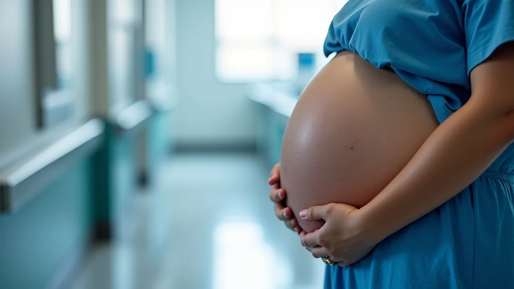
    Medical Check-Ups and Tests

    The second trimester is a key period for medical monitoring. Regular appointments with your healthcare provider are essential. These check-ups ensure both you and your baby are healthy and developing as expected.

    Several important tests are typically performed during these months.

    Routine Prenatal Visits

    During the second trimester, your prenatal appointments usually become more frequent. They might be scheduled every four weeks. These visits are crucial for tracking your progress and your baby’s growth.

    • Your healthcare provider will monitor your weight gain and blood pressure.
    • They will listen to your baby’s heartbeat using a Doppler device.
    • Your uterus size will be measured to ensure appropriate growth.

    These regular check-ins allow your provider to catch any potential issues early. They also provide a consistent opportunity for you to ask questions and voice any concerns you might have about your pregnancy.

    Anatomy Scan

    One of the most anticipated appointments in the second trimester is the anatomy scan, also known as the mid-pregnancy ultrasound. This detailed ultrasound is usually done between weeks 18 and 22. It allows for a thorough examination of your baby’s development.

    • The anatomy scan checks for the physical development of your baby’s organs and limbs.
    • It helps determine the baby’s sex, if you wish to know.
    • This scan can also identify potential birth defects or complications.

    Seeing your baby in such detail is a memorable experience for many parents-to-be. The information gathered from this scan is vital for confirming the baby’s health and planning for birth and beyond.

    Glucose Screening Test

    Around weeks 24 to 28, you will likely be offered a glucose screening test. This test is performed to check for gestational diabetes, a type of diabetes that can develop during pregnancy.

    • The test involves drinking a sugary solution and having your blood sugar levels measured after one hour.
    • If the results are high, a follow-up glucose tolerance test may be recommended.
    • Gestational diabetes needs to be managed to ensure a healthy pregnancy for both mother and baby.

    Early detection and management of gestational diabetes are important. It can help prevent complications such as preeclampsia or a larger-than-average baby. Your healthcare provider will guide you on the best course of action if this condition is diagnosed.

    Baby’s Growth and Development

    The second trimester is a time of incredible growth and development for your baby. They transform from a tiny fetus into a recognizable infant. Many of the major organs are formed and begin to function.

    This phase is critical for their maturation.

    Organ Development

    By the start of the second trimester, most of your baby’s basic body structure is in place. During these months, these structures mature and start working. The digestive system develops, and the baby begins to swallow amniotic fluid.

    • The baby’s skin starts to develop, and they begin to grow hair.
    • Lanugo, a fine downy hair, covers the body, and vernix caseosa, a waxy coating, protects the skin.
    • The lungs are developing but are not yet mature enough to function outside the womb.

    These internal developments are crucial for the baby’s survival and health after birth. The formation of these systems prepares them for life outside the uterus, though significant development continues.

    Sensory Development

    Your baby’s senses also start to develop and become more active during the second trimester. This means they are becoming more aware of their surroundings, both inside and outside the womb.

    • The baby can begin to hear sounds from the outside world, like your voice or music.
    • They can start to see, although their eyelids may still be closed for much of this period.
    • Taste and touch are also developing, with the baby reacting to the amniotic fluid and sensations.

    This sensory development is the beginning of your baby’s interaction with the world. Responding to these developing senses, like talking to your belly, can help create a bond even before birth.
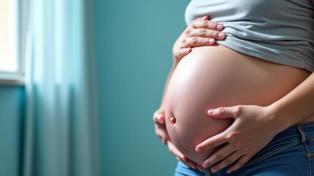
    Movement and Kicks

    A very exciting milestone in the second trimester is feeling your baby move, often referred to as “quickening.” As the baby grows, their movements become stronger and more noticeable.

    • You might first feel subtle flutters or bubbles, which grow into more distinct kicks and rolls.
    • These movements indicate healthy muscle development and nerve function.
    • By the end of the second trimester, the baby’s movements are often quite vigorous.

    Feeling these kicks is a profound experience. It’s a tangible connection to the life growing inside you and a reassuring sign of your baby’s well-being. Tracking these movements can become a cherished part of your pregnancy routine.

    Staying Healthy and Comfortable

    The second trimester is a great time to focus on maintaining a healthy lifestyle. With energy levels generally higher and fewer serious discomforts, you can actively work on your well-being. This helps support your baby’s growth and prepares you for the later stages of pregnancy.

    Nutrition and Hydration

    Proper nutrition is vital for both you and your baby. Your body needs extra nutrients to support fetal development and your own changing needs.

    • Focus on a balanced diet rich in fruits, vegetables, lean proteins, and whole grains.
    • Ensure adequate intake of key nutrients like folate, iron, calcium, and vitamin D.
    • Stay well-hydrated by drinking plenty of water throughout the day.

    A healthy diet provides the building blocks for your baby’s development. It also helps you maintain energy levels and prevent common pregnancy issues like anemia. If you’re unsure about your diet, consult with your healthcare provider or a registered dietitian.

    Exercise and Activity

    Continuing or starting gentle exercise can be highly beneficial during the second trimester. It helps manage weight gain, reduces aches and pains, and improves mood and sleep.

    • Low-impact exercises like walking, swimming, and prenatal yoga are excellent choices.
    • Listen to your body and avoid overexertion.
    • Always consult your healthcare provider before starting any new exercise program.

    Regular physical activity can make your body stronger and more resilient for labor and delivery. It also helps your body recover more easily postpartum. Finding an activity you enjoy makes it easier to stick with a routine.

    Rest and Sleep

    Although energy levels often improve, getting enough rest remains important. Your body is still working hard to grow your baby. Sleep can become more challenging as your pregnancy progresses.

    • Try to get 7-9 hours of sleep per night.
    • Establish a relaxing bedtime routine to help you wind down.
    • Use pillows to support your growing belly and back for a more comfortable sleep position, often on your side.

    Prioritizing sleep helps your body repair and recharge. It’s also crucial for your mental and emotional well-being. If you experience insomnia or frequent waking, discuss it with your healthcare provider.

    Common Myths Debunked

    Myth 1: You will feel your baby move very early in the second trimester

    The reality is that feeling your baby move for the first time, often called quickening, can vary greatly. For many first-time mothers, this sensation might not be noticeable until closer to the middle or even the end of the second trimester, around 18-24 weeks. Experienced mothers might feel it earlier.

    These early movements can be subtle, like faint flutters or gas bubbles, making them easy to miss at first.

    Myth 2: Morning sickness completely disappears by the second trimester

    While it’s true that many women experience a significant reduction or complete disappearance of morning sickness as they enter the second trimester, this isn’t universal. Some women continue to experience nausea and vomiting throughout their pregnancy, albeit often less severely. If morning sickness persists intensely, it’s important to discuss it with your healthcare provider, as it could be a sign of a condition like hyperemesis gravidarum.

    Myth 3: The second trimester is always the easiest part of pregnancy

    For many, the second trimester does offer increased energy and reduced nausea, making it feel easier. However, this period can also bring its own set of discomforts, such as backaches, heartburn, and swelling. For some individuals, pregnancy complications might also arise during this phase.

    Therefore, while often more comfortable, it’s not guaranteed to be universally “easy” for everyone.

    Myth 4: You must gain a specific amount of weight by the end of the second trimester

    Weight gain recommendations during pregnancy are personalized and depend on your pre-pregnancy weight and health. While there are general guidelines, there isn’t a single, strict number everyone must hit by the end of the second trimester. Healthcare providers assess weight gain based on individual needs.

    Focusing on balanced nutrition and healthy weight gain as advised by your doctor is more important than hitting an arbitrary number.

    Frequently Asked Questions

    Question: When do most women start showing their baby bump in the second trimester?

    Answer: Most women begin to show their baby bump more visibly between weeks 13 and 20 of the second trimester. First-time mothers might show a bit later than those who have been pregnant before.

    Question: Is it safe to travel during the second trimester?

    Answer: Generally, the second trimester is considered the safest time to travel during pregnancy for most healthy women. However, it’s always best to discuss any travel plans with your healthcare provider.

    Question: How much does the baby grow in the second trimester?

    Answer: The baby grows significantly, from about 3-4 inches and less than an ounce at the start to around 14-16 inches and 1.5-2 pounds by the end of the second trimester.

    Question: What should I do if I experience severe pain in my abdomen?

    Answer: Severe abdominal pain should always be reported to your healthcare provider immediately, as it can sometimes indicate a serious issue that needs prompt medical attention.

    Question: Can I feel my baby’s movements clearly by week 20?

    Answer: Many women can feel clear baby movements by week 20, especially if they have been pregnant before. However, it’s still within the normal range to feel them a bit later, and the strength and frequency of movements vary.

    Summary

    The second trimester is when many exciting developments occur. You’ll experience noticeable physical changes, your baby will grow rapidly, and important medical check-ups are scheduled. By focusing on good nutrition, exercise, and rest, you can feel your best during these months.

    You now have a clear picture of what this important phase of pregnancy involves.

  • Second Trimester Screening RACGP Guide

    Second Trimester Screening RACGP Guide

    Key Takeaways

    • You will learn what second trimester screening is all about.
    • We will explain the typical tests involved.
    • You’ll discover why this screening is important for expectant parents.
    • We’ll cover how the results are interpreted.
    • The post will help you understand the next steps after screening.
    • You’ll find out what to expect during the screening process.

    Understanding Second Trimester Screening RACGP

    Second trimester screening is a set of tests done when a baby is about 15 to 20 weeks old in the womb. These tests help doctors get more information about your baby’s health. They look for certain signs that might suggest a higher chance of specific conditions.

    It’s a way to get a clearer picture of your baby’s development and well-being during this important stage of pregnancy.

    What is Second Trimester Screening?

    This screening involves a combination of tests. These tests are not meant to diagnose a condition but rather to assess the risk. They help parents and doctors make informed decisions about further care.

    The timing is specific because the baby’s organs are developing, making them visible on ultrasound. Also, certain hormone levels in the mother’s blood are best measured during this period.

    Think of it like a detailed check-up for your growing baby. It’s not a definitive answer, but it gives valuable insights. These insights are based on scientific methods and data collected over many years.

    The aim is to provide expectant parents with information to help them prepare or plan accordingly.

    Why is it Done?

    The main reason for second trimester screening is to check for certain birth differences. These can include problems with the baby’s brain, heart, or other organs. It also screens for genetic conditions like Down syndrome and trisomy 18.

    Knowing about these possibilities early can make a big difference in how care is managed during pregnancy and after birth.

    The results can guide discussions about further testing if needed. It also helps healthcare providers prepare for any special care the baby might require right after delivery. For many parents, having this information offers peace of mind, regardless of the outcome.

    It’s about empowerment through knowledge.

    The RACGP Context

    The Royal Australian College of General Practitioners (RACGP) provides guidelines and resources for healthcare professionals in Australia. When we talk about second trimester screening racgp, it refers to the standards and recommendations set by this college for how GPs should discuss, order, and interpret these screenings for their patients. It ensures a consistent and high standard of care across the country.

    GPs play a key role in guiding expectant mothers through the options available. They explain what the tests involve, what the results mean, and what the next steps could be. The RACGP guidelines ensure that GPs are well-equipped to offer this vital service.

    They emphasize clear communication and patient autonomy.

    Components of Second Trimester Screening

    Second trimester screening typically involves two main parts: a blood test and an ultrasound. These tests work together to provide a more comprehensive risk assessment. Each part looks for different indicators that, when combined, offer a better picture than either test alone.

    The Maternal Blood Test

    This blood test measures the levels of certain substances in the mother’s blood. These substances, often called markers, can provide clues about the baby’s health. For example, specific hormone levels can be higher or lower if there’s an increased chance of certain genetic conditions.

    Common markers include Alpha-fetoprotein (AFP), human chorionic gonadotropin (hCG), and unconjugated estriol. Sometimes, inhibin-A is also measured. Changes in these levels don’t mean the baby definitely has a problem.

    Instead, they suggest a statistical possibility that requires further attention.

    Key Blood Markers Explained

    Alpha-fetoprotein (AFP) is a protein made by the baby’s liver. High levels in the mother’s blood might indicate a neural tube defect, like spina bifida. Low levels could sometimes be associated with Down syndrome.

    However, many factors can influence AFP levels, so it’s interpreted alongside other results.

    Human chorionic gonadotropin (hCG) is a hormone produced by the placenta. Higher levels are often seen in pregnancies with Down syndrome. Conversely, lower levels might be linked to trisomy 18.

    It’s a hormone that plays a vital role in maintaining pregnancy.

    Unconjugated estriol is another hormone produced by the placenta and the baby. Low levels of estriol can be associated with Down syndrome. It’s part of the complex hormonal balance during pregnancy.

    Inhibin-A is a hormone produced by the placenta. If measured, it’s often used with other markers to improve the accuracy of screening for Down syndrome. Elevated levels of inhibin-A are sometimes seen in pregnancies with Down syndrome.

    The Ultrasound Examination

    The ultrasound, often called a Level II or detailed anomaly scan, is a key part of the screening. This detailed scan allows a trained sonographer or radiologist to examine the baby’s anatomy very carefully. They look for physical signs that might suggest a genetic condition or developmental problem.

    During the ultrasound, measurements of the baby are taken. These include the head circumference, abdominal circumference, and femur length. The sonographer also visually checks the baby’s brain, heart, spine, limbs, and facial features.

    A specific measurement, the nuchal translucency (NT), is measured in the first trimester but can be reassessed in the second trimester. Other markers might also be looked for.

    Ultrasound Markers

    Several physical features can be assessed during the ultrasound that might indicate an increased risk. These are often referred to as sonographic markers. They are not definite proof of a condition but add to the overall risk assessment.

    Examples include a shortened nasal bone (hypoplastic nasal bone). This is often seen in babies with Down syndrome. Another marker is the presence of choroid plexus cysts in the baby’s brain.

    These are usually harmless but can sometimes be associated with trisomy 18.

    The scan also checks for things like the number of fingers and toes, the structure of the heart chambers, and the presence of the stomach. Any observed abnormalities are carefully documented and discussed with the healthcare team.

    Combining the Results

    The power of second trimester screening comes from combining the information from both the blood test and the ultrasound. A special computer program uses all this data, along with the mother’s age and family history, to calculate a specific risk number. This number represents the chance of the baby having a particular condition.

    For instance, the risk of Down syndrome might be reported as 1 in 100 or 1 in 5000. A higher number, like 1 in 100, means a greater chance. A lower number, like 1 in 5000, means a very low chance.

    It’s important to remember these are risk assessments, not diagnoses.

    Interpreting the Results

    Understanding the results of second trimester screening is crucial for expectant parents. The outcome will either indicate a low risk or a high risk for certain conditions. This distinction guides the next steps in pregnancy care.

    Low Risk Results

    If your screening results show a low risk, it means the chance of your baby having one of the tested conditions is very small. For example, a result of 1 in 5000 or less is generally considered low risk for Down syndrome. In most cases, no further testing for these specific conditions is recommended.

    This outcome often brings relief and allows parents to continue their pregnancy with greater reassurance. However, it’s still important to remember that screening tests are not perfect. There’s always a small chance of a false negative, meaning a condition might still be present even with a low-risk result.

    High Risk Results

    A high-risk result means that the screening tests have identified an increased chance of your baby having a genetic or developmental condition. For example, a risk of 1 in 100 for Down syndrome would be considered high risk. It’s vital not to panic if you receive a high-risk result.

    This result does not mean your baby definitely has the condition. It simply means the statistical likelihood is higher than in the general population. Your healthcare provider will discuss these results with you in detail and explain the options for further investigation.

    This often involves more definitive diagnostic tests.

    What are Diagnostic Tests?

    When screening results indicate a higher risk, diagnostic tests are usually offered. These tests can confirm whether the baby actually has a specific condition. The two main types of diagnostic tests are amniocentesis and chorionic villus sampling (CVS).

    Amniocentesis involves taking a small sample of the amniotic fluid that surrounds the baby. This fluid contains baby’s cells that can be tested for genetic abnormalities. CVS involves taking a small sample of placental tissue.

    Both procedures carry a small risk of miscarriage.

    Amniocentesis

    Amniocentesis is typically performed between 15 and 20 weeks of pregnancy, though it can be done later. A thin needle is guided through the mother’s abdomen into the amniotic sac. The fluid is withdrawn and sent to a lab for genetic analysis.

    The results usually take a week or two.

    This test can detect chromosomal abnormalities like Down syndrome, Edwards syndrome (trisomy 18), and Patau syndrome (trisomy 13). It can also identify certain single-gene disorders and neural tube defects. It provides a definitive diagnosis.

    Chorionic Villus Sampling (CVS)

    CVS is usually performed earlier in pregnancy, typically between 10 and 13 weeks. A sample of chorionic villi, which are finger-like projections from the placenta, is taken. This can be done either through the cervix (transcervical CVS) or through the abdomen (transabdominal CVS).

    Like amniocentesis, CVS can identify chromosomal abnormalities and some genetic disorders. It is generally faster than amniocentesis, with results available in about a week. Both procedures are invasive and have a small associated risk of complications.

    The Role of General Practitioners

    General Practitioners (GPs) are often the first point of contact for expectant parents regarding prenatal screening. They play a vital role in educating patients, facilitating the screening process, and providing support. The RACGP guidelines outline the expected responsibilities of GPs in this area.

    Discussing Options with Patients

    GPs are responsible for explaining the different prenatal screening and diagnostic options available to pregnant individuals. This includes the purpose of second trimester screening, its benefits, limitations, and potential risks. They ensure patients understand that screening is optional and provide unbiased information.

    The conversation should cover what each test entails, when they are performed, and what the results mean. This allows individuals to make informed decisions that align with their personal values and beliefs. It’s a dialogue, not just a prescription.

    Referral and Coordination

    Once a patient decides to proceed with second trimester screening, their GP will facilitate the necessary referrals. This includes referring them to a pathology laboratory for the blood test and to a radiology or ultrasound clinic for the scan. GPs ensure that the correct forms are completed and that the tests are ordered appropriately.

    They also play a role in coordinating the different parts of the screening process. This ensures that the blood sample is taken at the right time relative to the ultrasound. Effective coordination minimizes delays and maximizes the accuracy of the results.

    It is a key part of their duty of care.

    Follow-up and Support

    After the screening results are available, GPs are responsible for discussing them with the patient. If the results are low risk, they provide reassurance. If the results are high risk, they offer support and explain the next steps, including referrals to specialists or genetic counselors.

    This follow-up is crucial for managing patient anxiety and ensuring they receive appropriate care. GPs act as a central point of contact, helping patients navigate the sometimes complex healthcare system. Their ongoing support is invaluable throughout the pregnancy.

    Common Myths Debunked

    Myth 1: Second trimester screening can definitively tell me if my baby has a condition.

    Reality: Second trimester screening is a risk assessment tool, not a diagnostic test. It calculates the likelihood of certain conditions based on your baby’s development and your blood markers. It can tell you if your risk is high or low, but it cannot provide a definitive “yes” or “no” answer for most conditions.

    Diagnostic tests like amniocentesis or CVS are needed for a definite diagnosis.

    Myth 2: If I have a low-risk result, my baby will be perfectly healthy.

    Reality: A low-risk result significantly reduces the chance of the baby having the conditions screened for, but it does not eliminate it entirely. There’s a small chance of a false negative result, where the screening indicates a low risk, but the baby does have a condition. It’s important to continue with routine prenatal care and be aware of any signs or symptoms that may arise.

    Myth 3: Second trimester screening is only for older mothers.

    Reality: While the risk of certain chromosomal conditions like Down syndrome increases with maternal age, second trimester screening is offered to all pregnant individuals, regardless of age. Younger mothers can also have babies with these conditions, and screening provides valuable information for everyone. The risk assessment considers age, but other factors also contribute to the final risk calculation.

    Myth 4: The ultrasound part of the screening can harm my baby.

    Reality: Ultrasound technology uses sound waves to create images and has been used in pregnancy for decades. It is considered a safe diagnostic tool for both mother and baby when used by trained professionals according to established guidelines. There is no evidence to suggest that diagnostic ultrasounds performed during pregnancy cause harm to the developing baby.

    Frequently Asked Questions

    Question: When exactly is the second trimester?

    Answer: The second trimester of pregnancy typically spans from week 13 to week 28 of gestation. Second trimester screening tests are usually performed between 15 and 20 weeks of pregnancy.

    Question: What conditions does second trimester screening look for?

    Answer: It primarily screens for chromosomal abnormalities like Down syndrome (Trisomy 21), Edwards syndrome (Trisomy 18), and Patau syndrome (Trisomy 13). It can also help detect neural tube defects such as spina bifida.

    Question: Is second trimester screening mandatory?

    Answer: No, second trimester screening is optional. It is a personal choice for expectant parents, and healthcare providers will discuss the benefits and limitations to help them make an informed decision.

    Question: How accurate is second trimester screening?

    Answer: The accuracy varies depending on the specific tests used. Generally, combined screening in the second trimester can detect about 75-85% of Down syndrome cases. However, it can also produce false positives, where a high-risk result does not mean the baby has the condition.

    Question: What happens if my screening results are abnormal?

    Answer: An abnormal or high-risk screening result means your chance of having a baby with a specific condition is higher. Your healthcare provider will discuss this with you and offer diagnostic tests, such as amniocentesis or CVS, to get a definitive diagnosis.

    Final Thoughts

    Understanding second trimester screening racgp means knowing it’s a valuable tool for expectant parents. It helps assess risks for certain baby conditions. The process involves a blood test and an ultrasound.

    Your GP is key to explaining options and next steps. This screening provides information to support informed decisions about your pregnancy care.

  • Pregnancy Test Very Faint Second Line Explained

    Pregnancy Test Very Faint Second Line Explained

    Seeing a very faint second line on a pregnancy test can be confusing. Many people wonder if it means they are pregnant or if it’s just a false positive. This is a common experience, especially when you’re trying to get pregnant for the first time.

    Don’t worry, we’ll break down what that little line really means. We will go step-by-step so it’s super easy to understand. Get ready to learn what this test result tells you and what to do next.

    Key Takeaways

    • A faint second line usually indicates pregnancy.
    • Hormone levels (hCG) determine line darkness.
    • Testing too early can cause faint lines.
    • Different test brands have varying sensitivities.
    • Confirming with a doctor is important.
    • Retesting after a few days can clarify results.

    Understanding A Faint Second Line

    A pregnancy test works by detecting a hormone called human chorionic gonadotropin (hCG). This hormone is produced by the body shortly after a fertilized egg attaches to the uterine lining. The intensity of the lines on the test indicates the amount of hCG detected.

    A very faint second line means that hCG is present, but at a lower concentration. This is often seen in the early stages of pregnancy when hormone levels are just starting to rise.

    For beginners, interpreting a faint line can be tricky because they might expect a strong, clear second line. They might question its validity. This happens because hCG levels are still building up.

    The test is sensitive enough to pick up even small amounts. This early detection is a positive sign. It means the test is likely working correctly and picking up on early pregnancy signals.

    What Does hCG Mean

    Human chorionic gonadotropin (hCG) is a hormone that your body starts producing right after a fertilized egg implants in your uterus. Think of it as a signal that pregnancy has begun. This hormone is only present in significant amounts during pregnancy.

    Pregnancy tests are designed to detect this specific hormone in your urine or blood. The amount of hCG rises rapidly in the early weeks of pregnancy. This increase is what allows pregnancy tests to detect pregnancy.

    The way hCG levels change is key to understanding pregnancy tests. In the first few weeks, hCG doubles roughly every 48 to 72 hours. This rapid increase is why a test might show a faint line one day and a stronger line a few days later.

    The more hCG there is, the darker the positive line will appear on most tests. A faint line just means your hCG levels are at the lower end of the detection range.

    How Pregnancy Tests Work

    Pregnancy tests use a chemical reaction to detect hCG. They contain antibodies that bind to hCG molecules in your urine. When hCG is present, it triggers a color change.

    Most home pregnancy tests have a control line and a test line. The control line shows the test is working properly. The test line appears if hCG is detected.

    The test line might be faint if the hCG concentration is low.

    The sensitivity of a pregnancy test refers to how much hCG it can detect. Some tests are more sensitive than others. A more sensitive test can detect pregnancy earlier.

    This is why some tests might show a faint line sooner than others. The instructions on the test kit are very important. They tell you when to test and how to read the results.

    Following them closely helps ensure you get the most accurate reading possible.

    Why The Line Might Be Faint

    Several factors can contribute to a faint second line on a pregnancy test. The most common reason is testing too early. When you are in the very early stages of pregnancy, your hCG levels might be too low for the test line to appear dark.

    Another reason is the concentration of your urine. Testing first thing in the morning is usually recommended because your urine is more concentrated, meaning it has a higher level of hCG if you are pregnant.

    The type of test you use can also play a role. Different brands have different sensitivities. Some might show a faint line at lower hCG levels than others.

    Diluted urine, perhaps from drinking a lot of fluids before testing, can also lead to a fainter line. It is important to remember that even a faint line is usually a positive result. It indicates the presence of hCG.

    Interpreting Your Result

    When you see that faint second line, it’s natural to feel a mix of excitement and uncertainty. The most important thing to remember is that most pregnancy tests are designed to be highly accurate. If you followed the instructions and a second line, no matter how faint, has appeared within the specified time frame, it is very likely a positive result.

    This means your body is producing hCG, which is the key indicator of pregnancy.

    However, it is crucial to understand that the darkness of the line does not typically indicate the health or viability of the pregnancy. A faint line simply means that the hCG levels are present but not yet at a high concentration. This is completely normal in the early days and weeks of pregnancy.

    The key is that the line is there. It is not an evaporation line, which usually appears after the test reading time has passed and is colorless or gray.

    Timing Is Everything

    The timing of when you take a pregnancy test is one of the biggest factors in how clear the second line will be. Most home pregnancy tests are most accurate when taken after you miss your period. If you test too early, before your hCG levels have risen sufficiently, you might get a faint positive or even a false negative.

    This is because the fertilized egg may have only recently implanted, and hCG production is just beginning.

    For example, testing just a few days after ovulation might be too soon. Even if conception occurred, there hasn’t been enough time for hCG to build up to detectable levels. This is why many experts recommend waiting until at least the first day of your missed period to test.

    Some highly sensitive tests claim to detect pregnancy a few days before a missed period, but these are still best used when hCG levels are more established to avoid faint lines or inconclusive results.

    Evaporation Lines vs. Positive Lines

    One common point of confusion with faint lines is differentiating between a positive result and an evaporation line. Evaporation lines are faint, colorless or grayish lines that can appear on a pregnancy test after the test has already been read, usually outside the recommended time frame. They occur when urine evaporates from the test strip, leaving behind the chemical reaction without the presence of hCG.

    A true positive line, even if faint, should typically appear within the time limit stated in the test’s instructions and should have some color to it.

    To avoid confusion, always read the test within the specified time frame. If a line appears after this window, it is generally not considered a positive result. It’s also helpful to use tests that have a clear indicator for positive results, like a plus sign or a distinct second line, rather than relying on subtle color changes.

    If you are unsure, it is always best to retest after a few days or consult with a healthcare professional.

    What If The Line Is Barely Visible

    If the second line is barely visible or looks like a shadow, it is still usually considered a positive result. The important factor is that the line is present and has color. It signifies that hCG is being detected.

    Do not dismiss it simply because it is faint. Many women have conceived with faint positive lines on their tests.

    This situation often calls for retesting. Waiting a couple of days and testing again, ideally with first-morning urine, can provide a clearer result. If the line gets darker, it confirms that your hCG levels are rising, which is a strong indicator of ongoing pregnancy.

    If the line remains faint or disappears, it might suggest a chemical pregnancy or a false positive, though the latter is less common with modern tests.

    Next Steps After A Faint Line

    So, you’ve seen that faint second line and are wondering what to do next. The best approach is to confirm the result and monitor your body’s changes. The initial faint line is a sign that something is happening, and further steps will help clarify the situation.

    It’s a moment for patience and careful observation, rather than immediate alarm or celebration without confirmation.

    Your next actions will involve retesting and seeking professional medical advice. These steps are designed to give you a definitive answer and ensure you get the care you need if you are indeed pregnant. It is also a good time to start thinking about your health and what lifestyle changes might be beneficial for a potential pregnancy.

    Retesting For Clarity

    Since a faint line can be due to low hCG levels, retesting is a crucial step. Wait at least 24 to 48 hours before taking another test. During this time, your hCG levels should have increased, potentially resulting in a darker, more visible second line.

    Using your first-morning urine is highly recommended for retesting, as it is the most concentrated and will contain the highest level of hCG, making detection easier.

    When retesting, you can use the same brand of test or try a different one. Some people find that using a digital pregnancy test can offer a more straightforward answer, as they display words like “Pregnant” or “Not Pregnant” instead of relying on line interpretation. Remember to always follow the instructions on the test kit carefully.

    Seeing a progressively darker line over a few days is a very strong indicator of a healthy, ongoing pregnancy.

    Consulting A Healthcare Provider

    Regardless of the clarity of your pregnancy test result, it is always a good idea to consult with a healthcare provider, such as a doctor or a midwife. They can perform a blood test, which is more sensitive than urine tests and can detect hCG levels earlier and more accurately. A blood test can confirm pregnancy and give you a more precise reading of your hCG levels.

    Your healthcare provider will also be able to discuss your next steps, such as scheduling your first prenatal appointment. They can offer guidance on prenatal vitamins, nutrition, and any necessary lifestyle adjustments. Building a relationship with a healthcare provider early in your pregnancy journey is essential for a healthy outcome for both you and your baby.

    Tracking Symptoms

    While waiting for retesting or your doctor’s appointment, pay attention to any early pregnancy symptoms you might be experiencing. These can include fatigue, nausea (morning sickness), tender breasts, frequent urination, and food aversions or cravings. While these symptoms can sometimes be mistaken for premenstrual symptoms, their presence, combined with a faint positive pregnancy test, can be a good indicator that you are pregnant.

    However, it’s important to remember that not everyone experiences noticeable symptoms, especially in the very early stages. Conversely, some symptoms can occur for reasons other than pregnancy. Therefore, symptoms alone are not a definitive confirmation.

    They should be considered alongside the results of pregnancy tests and professional medical advice. Keep a journal of your symptoms and any changes you notice.

    Factors Affecting Test Accuracy

    While modern pregnancy tests are highly reliable, certain factors can influence their accuracy and potentially lead to a faint line or an unclear result. Understanding these factors can help you interpret your test results with greater confidence and know when to seek further clarification. It’s not just about having the hormone; it’s about how and when the test detects it.

    These elements can range from how the test is administered to the specific type of test being used. By being aware of them, you can maximize your chances of getting an accurate reading the first time, or at least understand why a result might be less clear than expected.

    Expiration Dates and Test Quality

    Like many products, pregnancy tests have expiration dates. Using a test that has passed its expiration date can lead to inaccurate results. The chemicals used in the test strip can degrade over time, making them less sensitive or entirely non-functional.

    Always check the expiration date on the packaging before using a test. If it’s expired, it’s best to discard it and purchase a new one.

    The quality and brand of the pregnancy test can also matter. While most reputable brands are reliable, there can be slight variations in sensitivity and performance. If you get an unclear result with one brand, it might be worth trying a different, well-regarded brand.

    Sometimes, a slightly more sensitive test might pick up hCG more clearly, even at low levels.

    Medications and Medical Conditions

    Certain medications can interfere with pregnancy test results. For instance, some fertility treatments contain hCG, which can cause a false positive result if you test too soon after administration. If you are undergoing fertility treatments, it’s crucial to discuss the timing of pregnancy testing with your doctor.

    They will advise you on when to test to get an accurate result, taking into account the hCG medication.

    Rarely, certain medical conditions can also affect test results. For example, a molar pregnancy or certain tumors can produce abnormally high levels of hCG, leading to very strong positive results. Conversely, some rare conditions or very early miscarriages might lead to rapidly dropping hCG levels, which could result in confusing or faint lines.

    If you have any concerns about underlying medical conditions, it’s important to discuss them with your healthcare provider.

    Pregnancy Test Very Faint Second Line Explained

    When you see a pregnancy test very faint second line, it typically means hCG is present in your urine, but at a low concentration. This is most common in the very early stages of pregnancy, when hCG levels are just beginning to rise. The test is sensitive enough to detect even small amounts of this pregnancy hormone.

    Therefore, that faint line, even if it’s barely visible, is usually a sign that you are pregnant.

    It’s important not to dismiss a faint line. Instead, consider it a signal to take further steps for confirmation. This includes retesting in a day or two, ideally with first-morning urine, and consulting with a healthcare professional.

    They can provide the most definitive confirmation through blood tests and guide you on the next steps in your pregnancy journey. This early detection is valuable.

    What Does A Faint Line Indicate

    A faint second line on a pregnancy test indicates a low level of human chorionic gonadotropin (hCG) in your urine. This hormone is produced by the body shortly after a fertilized egg implants in the uterus. The strength of the line generally correlates with the amount of hCG detected.

    A faint line means that the hCG levels are present but are still relatively low.

    This is very common in the early days of pregnancy. For instance, if you ovulated and conceived about a week ago, your hCG levels might be just high enough for a sensitive test to detect them. This doesn’t mean the pregnancy is less healthy; it simply means it is very early.

    It’s a sign that the process has begun, and the body is starting to signal pregnancy.

    Examples of Faint Line Scenarios

    Let’s look at some real-life scenarios where a faint second line might appear. Imagine Sarah, who just found out she’s one day late for her period. She takes a home pregnancy test in the afternoon, and a very faint line appears.

    Excited but cautious, she waits until the next morning and uses first-morning urine for another test. This time, the second line is a bit darker, confirming her pregnancy.

    Another example is Maria, who is experiencing early pregnancy symptoms like nausea. She decides to test a few days before her expected period. The test shows an extremely faint line.

    She retests a week later after her missed period, and the line is now very clear and dark. These examples show how early testing or low hCG levels can result in a faint line, which often becomes clearer with time.

    Understanding hCG Levels

    Human chorionic gonadotropin (hCG) levels are what pregnancy tests detect. These levels begin to rise soon after implantation. Implantation typically occurs about 6 to 12 days after fertilization.

    At the time of a missed period, hCG levels can range from about 25 mIU/mL to 100 mIU/mL in urine. A very faint line often indicates levels in the lower end of this range, perhaps around 25-50 mIU/mL.

    As pregnancy progresses, hCG levels double approximately every 48 to 72 hours. This rapid increase is why a faint line today might become a strong line in a few days. For instance, if your hCG is 50 mIU/mL today, in two days it could be around 100-200 mIU/mL.

    This increase in hormone concentration will make the test line much more visible.

    Comparing Test Sensitivity

    The sensitivity of a pregnancy test refers to the lowest level of hCG it can detect. Tests are usually measured in milli-international units per milliliter (mIU/mL). A highly sensitive test might detect hCG at levels as low as 10 mIU/mL, while less sensitive tests might require 50 mIU/mL or more to show a positive result.

    If you are using a less sensitive test very early on, you might get a negative result or a very faint line even if you are pregnant. Conversely, a highly sensitive test might detect pregnancy a few days earlier, but the line might be faint. Here’s a simple comparison of sensitivity levels:

    Test Sensitivity (mIU/mL) Likely Result Timing Typical Line Appearance
    10-20 mIU/mL Up to 5 days before missed period Potentially faint line
    25 mIU/mL Around missed period Visible line, possibly faint
    50+ mIU/mL After missed period Clear, darker line

    Choosing a test with a sensitivity of around 25 mIU/mL is often a good balance for detecting pregnancy around the time of your missed period, which is when hCG levels are usually sufficient for a clear result. If you are testing earlier, a test with lower mIU/mL detection is advisable, but expect a fainter line.

    Common Myths Debunked

    Myth 1: A faint second line always means a chemical pregnancy

    This is not true. While a faint line can sometimes indicate a chemical pregnancy (a very early miscarriage), it is much more commonly a sign of a healthy, ongoing pregnancy in its very early stages. Chemical pregnancies occur when an egg is fertilized and implants, but the pregnancy doesn’t continue to grow.

    However, many women who see a faint line go on to have perfectly normal pregnancies. The faintness is usually due to low hCG levels.

    Myth 2: Only a dark second line means you’re pregnant

    This is a common misconception. Pregnancy tests are designed to detect the presence of hCG. Any visible second line, no matter how faint, that appears within the recommended testing time and has color, usually indicates a positive result.

    The darkness of the line reflects the concentration of hCG, which naturally increases as the pregnancy progresses. A faint line simply means the concentration is currently low.

    Myth 3: You can tell the sex of the baby by the darkness of the line

    There is absolutely no scientific basis for this claim. The darkness of the second line on a pregnancy test is determined by the level of hCG in your urine. It has no connection to the sex of the fetus.

    The sex of the baby is determined by the chromosomes of the sperm that fertilizes the egg, not by the hCG levels. This myth is purely folklore.

    Myth 4: If the line is faint, the pregnancy won’t last

    This is another myth that causes unnecessary worry. A faint line primarily indicates that hCG levels are low because the pregnancy is very new. As the pregnancy develops, hCG levels rise, and subsequent tests will show darker lines.

    Many women have had successful pregnancies after seeing faint positive lines on their initial tests. It is the progression of hCG levels and confirmation from a doctor that matter most.

    Frequently Asked Questions

    Question: Can a faint second line be a false positive

    Answer: False positives are rare with modern pregnancy tests. A faint second line is almost always a true positive, indicating the presence of hCG. If you are concerned, retesting after a few days or seeing a doctor can confirm the result.

    Question: How soon can I retest if I see a faint line

    Answer: It’s generally recommended to wait at least 24 to 48 hours before retesting. This allows your hCG levels to increase, which may result in a darker, clearer line.

    Question: Should I use first-morning urine for retesting

    Answer: Yes, using first-morning urine is highly recommended. It is the most concentrated, meaning it will have the highest level of hCG if you are pregnant, making detection easier and potentially yielding a clearer result.

    Question: What if the faint line disappears after the test reading time

    Answer: If the line appears after the test’s specified reading window and has no color or is faint and colorless, it’s likely an evaporation line and not a positive result. Always read the test within the time frame indicated in the instructions.

    Question: What is the difference between a faint positive and an evaporation line

    Answer: A faint positive line is a colored line that appears within the test’s reaction time and indicates hCG presence. An evaporation line is typically colorless or grayish and appears after the test reading time as urine evaporates from the strip.

    Final Thoughts

    Seeing a pregnancy test very faint second line is often the first sign of pregnancy. It means hCG is present, even if at low levels. This is normal in early pregnancy.

    Retest in a couple of days with first-morning urine for clarity. Confirm with your doctor for peace of mind and the best care.

  • Understanding Second Trimester Loss RCOG

    Understanding Second Trimester Loss RCOG

    Losing a baby in the second trimester can be incredibly hard. For many, this is a topic that brings up many questions and worries. It might feel confusing because it happens later than very early losses but is still before viability.

    This post aims to make things clearer. We will walk through what you need to know step by step so you can feel more informed and prepared. Let’s explore what the RCOG guidelines say about second trimester loss rcog.

    Key Takeaways

    • You will learn about the definition and common causes of second trimester loss.
    • Understand the emotional and physical impact of this type of loss.
    • Discover what the RCOG guidelines recommend for care and support.
    • Find out about the steps involved in investigations after a loss.
    • Learn about resources and support systems available to you.

    What Is Second Trimester Loss

    Defining Second Trimester Loss According to RCOG

    The second trimester typically spans from week 13 to week 28 of pregnancy. A loss occurring within this period is medically referred to as a late miscarriage or a fetal loss. The Royal College of Obstetricians and Gynaecologists, or RCOG, provides guidelines to ensure consistent and high-quality care for individuals experiencing such losses.

    Understanding these definitions is the first step in grasping the scope of care and support available.

    The RCOG’s guidance helps healthcare professionals manage these sensitive situations. It ensures that both the medical aspects and the emotional needs of patients are addressed appropriately. This standardized approach aims to reduce uncertainty and improve the experience for those going through a very difficult time.

    Timing of Second Trimester Loss

    The second trimester is a crucial period where many significant fetal developments occur. A loss in this phase means that the fetus has developed past the early stages of pregnancy. This can make the experience feel particularly impactful for expectant parents.

    The RCOG clearly defines this period. The guidelines cover losses from the start of the 13th week up to the end of the 28th week of gestation. This specific window helps in classifying the type of loss and determining the appropriate medical pathways for investigation and management.

    RCOG Classification of Pregnancy Loss

    The RCOG categorizes pregnancy losses for clarity and consistency in care. Early pregnancy loss typically occurs before 12 weeks. Second trimester loss, as we are discussing, falls between 12 weeks and 24 weeks gestation in some definitions, while other guidelines might extend this to 28 weeks.

    This classification is important for several reasons. It influences the type of medical investigations recommended. It also guides the support offered to families.

    Knowing the RCOG’s definitions helps patients understand the medical framework surrounding their experience.

    Distinguishing from Other Pregnancy Complications

    It is important to differentiate second trimester loss from other pregnancy complications. For instance, ectopic pregnancies and molar pregnancies occur very early. Preterm birth refers to the birth of a baby who is born too early, usually before 37 weeks of pregnancy, but often implies survival of the baby.

    Second trimester loss, in the context of RCOG guidelines, specifically addresses fetal demise or expulsion between the defined gestational ages. This distinction is vital for accurate diagnosis and appropriate medical and emotional support.

    Causes Of Second Trimester Loss

    Common Medical Reasons For Late Miscarriage

    Several factors can lead to a loss in the second trimester. These reasons can be complex and varied, impacting different pregnancies differently. The RCOG guidelines often mention a range of potential causes that healthcare providers investigate.

    Identifying the cause, if possible, can sometimes help with future pregnancies and provide closure. However, it is also common for a specific cause not to be found, which can be distressing. Healthcare teams work to provide the best possible care regardless of whether a cause is identified.

    Chromosomal Abnormalities

    While more common in the first trimester, chromosomal abnormalities can also contribute to losses in the second trimester. These are errors in the number or structure of chromosomes, which are the building blocks of our cells. They can prevent a baby from developing properly.

    These abnormalities are usually a random event and not inherited. They happen when the sperm or egg cell is formed, or very early after fertilization. Medical professionals use genetic testing on placental tissue or the fetus, if available, to help determine if this was a factor.

    Maternal Health Conditions

    Certain health conditions in the mother can increase the risk of pregnancy loss. These can include chronic illnesses that are not well-managed. Examples include uncontrolled diabetes, high blood pressure (hypertension), and autoimmune disorders like lupus.

    Effective management of these conditions before and during pregnancy is key. Healthcare providers will often work closely with specialists to optimize the mother’s health. This helps to reduce the risks associated with these underlying conditions.

    Infections

    Infections can play a significant role in second trimester losses. Certain bacterial, viral, or parasitic infections can affect the uterus, placenta, or the developing baby. These can sometimes lead to preterm labor or direct harm to the fetus.

    Examples include infections of the urinary tract, sexually transmitted infections, or even common infections like the flu if severe. RCOG guidelines emphasize screening for and treating infections promptly during pregnancy to safeguard the pregnancy.

    Problems With the Placenta

    The placenta is vital for supporting the baby’s growth and development. Issues with the placenta can unfortunately lead to pregnancy loss. These problems can involve the placenta not forming correctly or separating from the uterine wall too early (placental abruption).

    Conditions like pre-eclampsia, a serious condition involving high blood pressure in pregnancy, can also affect placental function. RCOG guidelines highlight the importance of monitoring placental health, especially in women with risk factors.

    Cervical Incompetence

    Cervical incompetence, also known as a weak cervix, means the cervix opens too early before a full-term pregnancy. This can cause the baby to be born too soon, leading to a second trimester loss. It often happens without pain or contractions.

    This condition can be diagnosed through ultrasound or based on a history of previous early losses. Treatment may involve a procedure to stitch the cervix closed during pregnancy, known as cervical cerclage, as recommended by RCOG standards.

    Emotional And Physical Impact

    The Grief Process After Loss

    Experiencing a second trimester loss is a profound grief. It is a deeply personal and often overwhelming experience. The grief can manifest in many ways, affecting a person’s emotions, thoughts, and behaviors.

    It’s important to acknowledge that there is no right or wrong way to grieve. Allowing yourself to feel a full range of emotions is a natural part of healing. Support from loved ones and professionals can be invaluable during this time.

    Navigating Emotional Challenges

    Feelings of sadness, anger, guilt, and confusion are common. Some individuals may also experience anxiety about future pregnancies or feel a sense of isolation. The RCOG acknowledges the significant psychological impact of pregnancy loss.

    It’s okay to seek professional help, such as counseling or therapy, to navigate these emotions. Talking about your feelings with a trusted friend, family member, or support group can also be very helpful. Remember, you are not alone in this.

    Physical Recovery

    After a second trimester loss, the body also needs time to heal. There can be physical symptoms that require attention and care. Medical professionals provide guidance on managing these aspects.

    This can include managing bleeding, pain, and hormonal changes. It is important to follow medical advice and attend any scheduled follow-up appointments to ensure a safe physical recovery. Rest and self-care are essential during this period.

    Support For Partners And Family

    It is vital to remember that partners and other family members also experience grief. Their feelings and needs are just as valid. Shared grief can strengthen bonds, but it can also create challenges if not addressed openly.

    Open communication between partners is key. Families can support each other by acknowledging their collective pain and offering comfort. Healthcare providers can also offer resources for partners and families.

    RCOG Guidelines On Care And Support

    Understanding The RCOG’s Approach

    The RCOG provides comprehensive guidelines for the management of pregnancy loss. These guidelines are designed to ensure that all individuals receive sensitive, respectful, and effective care. They cover all aspects, from diagnosis to follow-up.

    The RCOG’s recommendations emphasize a patient-centered approach. This means that decisions about care are made in partnership with the individual, considering their wishes and circumstances. The focus is on providing both medical expertise and emotional support.

    Medical Management Options

    Depending on the circumstances of the loss, there are different medical management options. These can include expectant management (waiting for the body to expel the pregnancy naturally), medical management (using medication to help the process), or surgical management (a procedure to remove the pregnancy tissue).

    The RCOG guidelines detail the criteria for each option and the associated risks and benefits. Healthcare providers discuss these choices thoroughly with patients to enable informed decision-making. This ensures that the chosen path aligns with the individual’s needs and preferences.

    Care During and After Loss

    The RCOG guidelines stress the importance of compassionate care throughout the entire process. This includes clear communication from healthcare professionals, pain management, and emotional support. After the loss, follow-up care is crucial.

    This follow-up may involve medical check-ups to ensure physical recovery. It also includes offering opportunities to discuss the loss, ask questions, and receive information about future pregnancies. Psychological support is also a key component of the RCOG’s recommendations.

    Investigations After Loss

    When a second trimester loss occurs, investigations may be recommended to try and determine the cause. This is not always possible, but it can be helpful for some individuals and for future family planning. The RCOG outlines a systematic approach to these investigations.

    These may include tests on the mother, such as blood tests for clotting disorders or infections. Investigations might also involve examining the placenta or, with consent, the fetus. The extent of investigations is usually tailored to the individual’s situation and wishes.

    Support Services Recommended by RCOG

    The RCOG strongly advocates for access to appropriate support services. This includes psychological support, counseling, and access to support groups. They recognize that grieving is a process that requires ongoing support.

    Healthcare providers are encouraged to inform individuals about these services. This ensures that those affected by pregnancy loss know where to turn for help. The goal is to provide holistic care that addresses both physical and emotional well-being.

    Investigations Following Second Trimester Loss

    Purpose of Investigations

    Investigating a second trimester loss is aimed at identifying any underlying causes. This information can be invaluable for several reasons. It can help healthcare providers offer more specific advice for future pregnancies.

    For some, knowing a cause can provide a sense of understanding and reduce uncertainty.

    The RCOG guidelines suggest a tailored approach to investigations. Not every loss will require the same level of investigation. The decision is usually made in discussion with the individual, considering their medical history and the circumstances of the loss.

    When Are Investigations Considered

    Investigations are typically considered after a second trimester loss. The decision to proceed often depends on factors such as the gestational age at the time of loss, whether there is a history of previous losses, and the clinical findings. The RCOG provides a framework for when these investigations might be most beneficial.

    If a specific risk factor is identified during the pregnancy, such as infection or a known maternal health condition, investigations will often focus on that area. Even without a clear risk factor, a general workup may be offered.

    Types of Investigations

    A range of investigations may be undertaken. These can involve tests on the mother, such as blood tests to check for clotting issues, infections, or hormonal imbalances. Imaging tests like ultrasounds may also be used to assess the uterus and ovaries.

    In some cases, if ethically appropriate and with full consent, investigations may extend to the fetus and placenta. This can include genetic testing to look for chromosomal abnormalities. The specific tests offered will be explained in detail by the healthcare team.

    Genetic Testing

    Genetic testing can be a crucial part of the investigation process. It helps to identify if a chromosomal abnormality in the fetus was the cause of the loss. This can provide significant information for future family planning.

    The RCOG guidelines support the offer of genetic testing where appropriate. This may involve testing tissue from the fetus or placenta. The process and implications of genetic testing are explained thoroughly to ensure informed consent.

    Maternal Blood Tests

    Blood tests on the mother are common investigations. These can help detect a variety of potential issues. For example, tests might check for thrombophilias, which are conditions that increase the risk of blood clots.

    Infections, such as those affecting the uterus or cervix, can also be identified.

    Hormonal levels might be assessed, particularly if there are concerns about conditions like thyroid disease. These blood tests are generally straightforward and provide valuable insights into maternal health that could impact pregnancy.

    Placental Examination

    Examining the placenta after a loss can offer important clues about why the pregnancy may not have continued. The placenta is a complex organ that nourishes the baby. Any abnormalities in its structure or function can have serious consequences.

    A pathologist can examine the placenta for signs of infection, inflammation, or problems with blood flow. This examination is a non-invasive way to gain understanding, especially when other investigations do not yield a clear answer.

    Coping And Support Resources

    Seeking Emotional Support

    Coping with the emotional aftermath of a second trimester loss is a journey. It is essential to have a strong support system in place. This can include loved ones, friends, and professional support services.

    The RCOG recognizes the profound grief associated with pregnancy loss and emphasizes the importance of psychological support. Allowing yourself to express your feelings is a vital part of healing.

    Professional Counseling And Therapy

    Many people find professional counseling or therapy extremely beneficial. Therapists trained in grief and loss can provide a safe space to explore complex emotions. They can offer coping strategies and help individuals process their experience.

    The RCOG encourages healthcare providers to refer individuals to mental health professionals. These services can help manage anxiety, depression, and trauma associated with pregnancy loss. There are various therapeutic approaches that can be helpful.

    Support Groups

    Connecting with others who have gone through similar experiences can be incredibly validating. Support groups, whether in-person or online, offer a sense of community and shared understanding.

    These groups provide a platform to share stories, fears, and hopes. Hearing from others who are on their own healing path can offer comfort and reduce feelings of isolation. Information on finding suitable support groups can often be obtained from healthcare providers.

    Information For Future Pregnancies

    For those planning future pregnancies, obtaining clear information and support is crucial. Healthcare providers can offer tailored advice based on previous pregnancy history and any identified causes of loss.

    The RCOG guidelines support comprehensive pre-conception counseling. This ensures that individuals are well-informed about risks, management strategies, and the support available for a subsequent pregnancy.

    Self-Care Strategies

    During the recovery period, prioritizing self-care is essential. This includes getting enough rest, eating nutritious food, and engaging in gentle physical activity when able. Allowing yourself time and space to grieve is also a form of self-care.

    Activities that bring comfort and peace, such as spending time in nature or engaging in hobbies, can be helpful. It is important to be patient with yourself and acknowledge that healing takes time.

    Common Myths Debunked

    Myth 1: Second trimester loss is always preventable.

    Reality: While some factors contributing to second trimester loss can be managed or prevented, many causes are not. Chromosomal abnormalities, for example, are often random events beyond anyone’s control. The RCOG guidelines focus on providing the best possible care and support when losses occur, rather than implying all losses are preventable.

    Myth 2: You should be able to get pregnant again quickly after a loss.

    Reality: While some individuals may conceive soon after a loss, emotional and physical recovery takes time. It is important to wait until you feel ready, both physically and emotionally. Healthcare providers usually recommend waiting a specific period, often until after a normal menstrual cycle, but individual circumstances vary.

    The RCOG encourages a personalized approach to planning future pregnancies.

    Myth 3: It’s your fault if you have a second trimester loss.

    Reality: This is a deeply harmful myth. In most cases, second trimester loss is not due to anything the pregnant person did or did not do. The causes are often medical and complex.

    The RCOG emphasizes that pregnancy loss is not a reflection of personal failure and that individuals deserve compassion and support.

    Myth 4: You don’t need medical follow-up after a second trimester loss.

    Reality: Medical follow-up is crucial after any pregnancy loss. Healthcare providers need to ensure your physical recovery is progressing well and address any potential complications. They also offer a space to discuss the loss, ask questions, and receive guidance for future pregnancies.

    RCOG guidelines strongly advocate for comprehensive follow-up care.

    Frequently Asked Questions

    Question: What is the main difference between first and second trimester loss according to RCOG?

    Answer: The main difference is the timing. First trimester loss typically occurs before 12 weeks of pregnancy, while second trimester loss, as defined by RCOG, occurs between 12 and 28 weeks of gestation.

    Question: Do RCOG guidelines cover what happens after the loss itself?

    Answer: Yes, RCOG guidelines extensively cover the period after a loss. They emphasize compassionate care, appropriate medical follow-up, and access to psychological support services for individuals and their families.

    Question: Is it always possible to find the cause of a second trimester loss?

    Answer: No, it is not always possible to find a definitive cause for a second trimester loss. Investigations are offered to help identify causes, but in many cases, a specific reason may remain unexplained.

    Question: Should I expect pain during a second trimester loss?

    Answer: Pain levels can vary. Some individuals experience cramping similar to menstrual pain, while others may experience more significant pain. Pain management is a key part of the care outlined in RCOG guidelines.

    Question: Where can I find support if I’ve experienced a second trimester loss?

    Answer: Support is available from healthcare providers, who can offer referrals to counselors and support groups. Many charities and organizations also provide specialized support for those who have experienced pregnancy loss.

    Conclusion

    Experiencing a second trimester loss is a deeply challenging event. The RCOG provides important guidelines to ensure you receive sensitive and comprehensive care. Understanding the causes, the impact, and the available support is key to navigating this difficult time.

    Remember that seeking help and allowing yourself to grieve are vital steps.

  • Third Trimester Pregnancy ICD 10 Codes Made Easy

    Third Trimester Pregnancy ICD 10 Codes Made Easy

    Getting the right codes for medical notes can feel tricky, especially when you’re learning. For anyone working with medical billing or records, understanding the third trimester of pregnancy icd 10 codes is super important. It might seem like a lot at first, but it doesn’t have to be hard.

    We’ll walk through this step by step, making it simple for you. Stick around, and you’ll be a pro at this in no time.

    Key Takeaways

    • You will learn how to correctly identify and use ICD-10 codes for the third trimester of pregnancy.
    • We will explore common reasons why pregnant individuals may need specific ICD-10 codes.
    • You will discover the primary ICD-10 codes used for routine third-trimester care.
    • The post will explain how to add codes for specific conditions that arise during this stage.
    • You will understand how to properly document and code for any follow-up care after delivery.
    • We will clarify common mistakes beginners make with these codes and how to avoid them.

    Understanding Third Trimester Pregnancy ICD-10 Codes

    This section is all about making sure you get a good grasp of why we use special codes for the third trimester of pregnancy. In medical coding, every little detail counts. ICD-10 codes are like a secret language that doctors and hospitals use to talk about illnesses and conditions.

    When a person is in their third trimester, which is from about week 28 until birth, certain things can happen that need to be noted. These codes help keep track of everything happening with the mom and the baby. They are used for billing, for research, and to make sure everyone taking care of the patient knows exactly what’s going on.

    For beginners, this might seem a bit confusing because there are many codes to remember. We will break down the most common ones and explain them in simple terms.

    What Are ICD-10 Codes?

    ICD-10 stands for the International Classification of Diseases, 10th Revision. It’s a system used worldwide to classify diseases, injuries, and causes of death. Think of it as a giant catalog of medical conditions.

    Each condition has a unique code, usually a mix of letters and numbers. This helps ensure that everyone is using the same terms and codes when talking about health issues. This consistency is vital for accurate record-keeping, billing, and statistical analysis.

    It allows health professionals to share information easily and understand patient histories without confusion.

    The ICD-10 system is very detailed. This detail is important because it allows for specific documentation of a patient’s health status. For instance, a simple cough might have one code, but a cough caused by pneumonia will have a different, more specific code.

    This level of detail is especially true for something as dynamic as pregnancy.

    Why Specific Codes for Pregnancy?

    Pregnancy is a unique and complex process. It involves significant changes in the mother’s body and the development of a new life. Because of this, a special chapter in the ICD-10 manual is dedicated to pregnancy, childbirth, and the postpartum period.

    This chapter, Chapter 15 (Pregnancy, Childbirth, and the Puerperium), uses codes that start with the letter ‘O’. These codes provide a standardized way to record all aspects of a pregnancy. This includes normal pregnancies, complications, and outcomes.

    Using specific pregnancy codes helps in tracking maternal and infant health trends. It allows healthcare providers to identify common issues faced by pregnant individuals. It also helps in planning for appropriate care and resources.

    For example, if many women in a certain area are experiencing gestational diabetes, this data can prompt public health initiatives.

    The Third Trimester Context

    The third trimester is the final stage of pregnancy, typically from week 28 to delivery. This period is often when the most significant fetal growth occurs and when mothers may experience more discomfort or specific health concerns. Common issues can include fatigue, back pain, swelling, and the baby getting ready for birth.

    Some women may also develop complications like preeclampsia or gestational diabetes.

    Because of these potential changes and issues, the third trimester requires careful monitoring. The ICD-10 codes reflect this by allowing for the documentation of a pregnancy’s progression and any deviations from the norm. Accurately coding this phase ensures that healthcare providers can manage care effectively and that data collected is precise.

    Common ICD-10 Codes for the Third Trimester

    This part focuses on the actual codes you’ll likely use when dealing with pregnant individuals in their third trimester. Remember, ICD-10 coding relies on specificity. This means you often need more than one code to fully describe a patient’s situation.

    We’ll cover codes for routine visits and then look at how to add codes for common conditions that might pop up. Getting these right is a big step towards accurate medical records.

    Routine Third Trimester Visits

    When a pregnant person comes in for a regular check-up during their third trimester, and everything is going smoothly, there’s a primary code to use. This code signifies a normal, healthy pregnancy. It’s important to select the correct code to show that there are no immediate concerns.

    These routine visits are crucial for monitoring the well-being of both the mother and the baby as the due date approaches.

    The most common code for a normal, current pregnancy is Z3A.xx, where ‘xx’ represents the specific week of gestation. For example, Z3A.28 would indicate 28 weeks of pregnancy. This code is always used in conjunction with another code to indicate the status of the pregnancy.

    For a normal, ongoing pregnancy without complications, you would typically use a code from the ‘O’ series.

    The primary code used to indicate that a patient is currently pregnant and receiving care is Z34.0, Supervision of Normal First Pregnancy, or Z34.9, Supervision of Unspecified Normal Pregnancy. When coding for a routine third-trimester visit, you would combine a code from the Z34.x series with the appropriate Z3A.xx code for the current week of gestation. This combination paints a clear picture of a healthy pregnancy at a specific stage.

    Coding for Specific Conditions

    Often, pregnant individuals in their third trimester may develop specific conditions that need to be coded. These can range from common discomforts to more serious health issues. Adding these codes provides a more complete medical history and ensures proper treatment and billing.

    It’s essential to be familiar with the codes for these common pregnancy-related conditions.

    For instance, if a pregnant person develops gestational hypertension, a specific code is used. This is distinct from preeclampsia or chronic hypertension. Similarly, conditions like anemia, urinary tract infections, or back pain during pregnancy all have their own ICD-10 codes.

    Being precise with these codes helps manage the patient’s care effectively.

    Examples of conditions and their associated codes include:

    • Gestational Hypertension (without significant proteinuria): O13.1
    • Pre-eclampsia, severe: O14.1
    • Anemia due to blood loss, third trimester: O99.013
    • Urinary tract infection, site not specified, pregnant, trimester 3: O99.533
    • Low back pain, third trimester: O99.843

    These codes, when used with the primary pregnancy code (like Z34.x and Z3A.xx), give a detailed account of the patient’s health status.

    Code Linking and Sequencing

    In ICD-10 coding, the order of codes often matters. Generally, the code that represents the primary reason for the encounter is listed first. For pregnancy-related encounters, the pregnancy code often takes precedence.

    However, this can change if a condition is so severe that it becomes the main focus of the visit.

    For example, if a patient is in their third trimester (Z34.x, Z3A.xx) and is admitted to the hospital for severe pre-eclampsia (O14.1), the pre-eclampsia code might be sequenced first on certain claims or reports, depending on specific payer guidelines. It’s always good practice to consult the official ICD-10-CM guidelines for the most accurate sequencing rules. These guidelines are updated annually and provide the definitive rules for coding.

    Understanding how to link and sequence codes correctly is vital for accurate reimbursement and for building a comprehensive patient record. It ensures that the full picture of the patient’s health is communicated. This practice prevents claim denials and ensures appropriate medical necessity is documented.

    Coding for Complications and Specific Trimester Issues

    This section delves deeper into coding for the issues that specifically arise or become more prominent in the third trimester. We will look at conditions that are common during this final phase of pregnancy and how to accurately represent them using ICD-10 codes. This knowledge is key for comprehensive documentation.

    Preeclampsia and Eclampsia

    Preeclampsia is a serious condition that can develop during pregnancy, typically after 20 weeks of gestation. It is characterized by high blood pressure and signs of damage to other organ systems, most often the liver and kidneys. Eclampsia is a more severe form of preeclampsia that includes seizures.

    The third trimester is a common time for preeclampsia to manifest or worsen.

    The ICD-10 codes for preeclampsia vary based on severity and whether it is associated with other conditions.

    • O14.0 Preeclampsia, mild
    • O14.1 Preeclampsia, severe
    • O14.2 HELLP syndrome (Hemolysis, Elevated Liver enzymes, Low Platelets)
    • O15.0 Eclampsia, unspecified
    • O15.1 Eclampsia, complicating pregnancy

    When coding for preeclampsia, it’s essential to use the most specific code available based on the physician’s documentation. For instance, if a patient has severe preeclampsia that is impacting their pregnancy, the code O14.1 would be used, along with the appropriate Z3A.xx code for their week of gestation.

    A statistic from the Centers for Disease Control and Prevention (CDC) shows that preeclampsia affects about 1 in 25 pregnancies in the United States. Early detection and proper coding are crucial for managing this potentially life-threatening condition.

    Gestational Diabetes Mellitus (GDM)

    Gestational diabetes is a type of diabetes that develops during pregnancy. It usually goes away after the baby is born. However, it can cause health problems for both the mother and the baby during pregnancy and delivery.

    The third trimester is when screening for GDM typically occurs, and if diagnosed, it requires specific coding.

    The primary ICD-10 code for Gestational Diabetes Mellitus is O24.41x. The last character indicates the trimester:

    • O24.411 Gestational diabetes mellitus in pregnancy, first trimester
    • O24.412 Gestational diabetes mellitus in pregnancy, second trimester
    • O24.413 Gestational diabetes mellitus in pregnancy, third trimester
    • O24.419 Gestational diabetes mellitus in pregnancy, unspecified trimester

    So, for a patient diagnosed with GDM in their third trimester, you would use O24.413. This code should be used in conjunction with the appropriate Z3A.xx code and any other relevant codes, such as those for managing high blood sugar or any complications arising from the GDM.

    Coding for GDM also requires noting if the patient requires insulin. If insulin is required, additional codes or specificity within the O24.413 code may be necessary depending on the detailed coding guidelines.

    Other Common Third Trimester Issues

    Beyond preeclampsia and gestational diabetes, several other issues commonly arise in the third trimester. These can include:

    • Edema in pregnancy: Swelling, especially in the legs and feet, is very common.
    • Back pain: As the baby grows, the mother’s posture changes, leading to back discomfort.
    • Heartburn and indigestion: The growing uterus can put pressure on the stomach.
    • Braxton Hicks contractions: These are ‘practice’ contractions that help prepare the body for labor.

    The ICD-10 codes for these issues are important for capturing the full patient experience. For example:

    • O26.833: Edema of legs, bilateral, complicating pregnancy, third trimester.
    • O99.893: Other specified diseases and conditions complicating pregnancy, childbirth and the puerperium, third trimester. This is a more general code that can be used for issues like back pain if a more specific code isn’t applicable.
    • K30: Functional dyspepsia, which can be used for severe indigestion.

    These codes help illustrate the day-to-day realities of pregnancy and ensure that all aspects of patient care are documented. They contribute to a complete medical picture, aiding in treatment and research.

    Coding for Fetal Issues and Monitoring

    Sometimes, concerns arise about the baby’s well-being during the third trimester. This can lead to increased monitoring. These situations also require specific ICD-10 codes.

    • O36.5 Maternal care for known or suspected fetal abnormality and damage, fetus affected.
    • O36.511 Maternal care for known or suspected malformation and/or defect of the central nervous system, fetus affected, first trimester. (Note: The trimester would change to 3 for the third trimester).
    • O36.6 Maternal care for excessive fetal growth.
    • O36.8 Maternal care for other specified fetal problems. This can include concerns about fetal movement or growth.

    When a fetus is identified with a condition that requires special care or monitoring, these codes are critical. They signal to other healthcare providers that there are specific concerns about the baby. This ensures that the delivery team is fully prepared.

    An example scenario: A routine ultrasound in the third trimester shows the baby is measuring larger than expected. The provider might use code O36.6 (Maternal care for excessive fetal growth) along with the appropriate Z3A.xx code. This alerts the care team to the potential need for managing macrosomia, which could affect delivery plans.

    Practical Application and Scenarios

    Understanding the codes is one thing, but knowing how to apply them in real-life situations is another. This section provides practical examples to help solidify your understanding of third trimester of pregnancy icd 10 coding. We will walk through a few common scenarios that coders might encounter.

    Scenario 1: Routine Check-up with Minor Complaint

    A 30-year-old patient, 32 weeks pregnant, comes in for a routine check-up. She reports mild swelling in her ankles but her blood pressure is normal and there are no other concerns for herself or the baby.

    1. Identify the primary reason for the visit: Supervision of a normal pregnancy.
    2. Determine the week of gestation: 32 weeks.
    3. Select the code for normal pregnancy supervision: Z34.0 (Supervision of Normal First Pregnancy) or Z34.9 (Supervision of Unspecified Normal Pregnancy). Since it’s her first pregnancy, Z34.0 is more specific.
    4. Add the code for the week of gestation: Z3A.32.
    5. Code the minor complaint: Edema of lower limbs complicating pregnancy. You would look for a code like O26.833 (Edema of legs, bilateral, complicating pregnancy, third trimester).

    The coded encounter might look like this: Z34.0, Z3A.32, O26.833. This clearly documents a healthy pregnancy at 32 weeks with a common, mild symptom.

    Scenario 2: Diagnosis of Gestational Diabetes

    A 28-year-old patient, 29 weeks pregnant, has just been diagnosed with Gestational Diabetes Mellitus after her glucose screening test. Her blood sugar levels are currently controlled with diet alone.

    1. Identify the primary diagnosis: Gestational Diabetes Mellitus.
    2. Determine the trimester: Third trimester.
    3. Select the code for GDM in the third trimester: O24.413.
    4. Determine the week of gestation: 29 weeks.
    5. Add the code for the week of gestation: Z3A.29.
    6. Add a code to indicate supervision of a normal pregnancy, as this is an added condition to a generally normal pregnancy at this stage: Z34.0 or Z34.9.

    The coded encounter might be: O24.413, Z3A.29, Z34.0. This shows a normal pregnancy at 29 weeks complicated by GDM managed with diet.

    Scenario 3: Labor and Delivery with Complication

    A patient arrives at the hospital in active labor at 38 weeks gestation. She has a history of severe preeclampsia, which is currently being monitored.

    1. Identify the main reason for admission and service: Labor and delivery.
    2. Determine the week of gestation: 38 weeks.
    3. Identify the significant complication: Severe preeclampsia.
    4. Select the primary code for the complication: O14.1 (Preeclampsia, severe).
    5. Select the code for the week of gestation: Z3A.38.
    6. Select a code for the stage of pregnancy and delivery. For delivery, codes starting with ‘O’ that indicate childbirth (e.g., O80 for normal spontaneous delivery, or more specific codes if operative delivery occurs) would be used, often sequenced after the complication.

    The coding here becomes more complex as it involves the outcome of the pregnancy. A typical sequencing might place the severe preeclampsia first if it’s the primary driver of medical decision-making, followed by the week of gestation and then the delivery code. For example: O14.1, Z3A.38, O80 (if it was a spontaneous vaginal delivery).

    Resources for Further Learning

    The ICD-10-CM Official Guidelines for Coding and Reporting are the definitive source for coding rules. These guidelines are updated annually and are essential for accurate coding. They can be found on the Centers for Disease Control and Prevention (CDC) website.

    Online coding resources and software also provide valuable tools. These can help verify codes and ensure they are current. Many professional organizations offer training and certification in medical coding.

    Common Myths Debunked

    Myth 1: All pregnancies are coded the same way.

    This is not true. While there are core codes for a normal pregnancy, the ICD-10 system is designed to be highly specific. Each pregnancy is unique, and coding needs to reflect any complications, individual patient factors, or specific trimester issues.

    Using a generic code for all pregnancies would miss vital health information.

    Myth 2: You only need one code per patient encounter.

    In most cases, especially during pregnancy, more than one code is needed. You typically need a code for the specific condition or reason for the visit, a code for the stage of pregnancy (week of gestation), and potentially codes for any co-existing conditions or symptoms. Accurate coding requires a complete picture.

    Myth 3: ICD-10 codes for pregnancy are only for the mother.

    While many codes focus on the mother’s health, ICD-10 also includes codes related to the fetus and newborn conditions. Codes like O36.x series, which relate to maternal care for fetal problems, directly address fetal well-being and are part of the pregnancy coding framework.

    Myth 4: The third trimester has only a few specific codes.

    The third trimester, like other stages of pregnancy, has a broad range of codes. These cover everything from normal supervision to specific complications like preeclampsia, gestational diabetes, fetal distress, and issues related to the mother’s health that arise during this critical final phase. The specificity allows for detailed medical record-keeping.

    Frequently Asked Questions

    Question: What is the main ICD-10 code for a normal third trimester pregnancy?

    Answer: The main codes used are Z3A.xx for the specific week of gestation and a code from the Z34.x series (like Z34.0 for first pregnancy or Z34.9 for unspecified normal pregnancy) to indicate supervision of a normal pregnancy.

    Question: Do I need a separate code for swelling in the legs during the third trimester?

    Answer: Yes, if edema is a noted issue, you would use a specific code like O26.833 (Edema of legs, bilateral, complicating pregnancy, third trimester) in addition to the pregnancy supervision codes.

    Question: How do I code for gestational diabetes in the third trimester?

    Answer: You would use code O24.413 for gestational diabetes mellitus in pregnancy, third trimester, along with the Z3A.xx code for the week of gestation.

    Question: What if the patient has multiple conditions in the third trimester?

    Answer: You would code each condition and reason for the encounter using the most specific ICD-10 codes available, sequencing them according to official coding guidelines.

    Question: Are there codes for monitoring fetal growth in the third trimester?

    Answer: Yes, codes such as O36.6 (Maternal care for excessive fetal growth) are used to document concerns and monitoring related to fetal development in the third trimester.

    Summary

    Coding for the third trimester of pregnancy involves using specific ICD-10 codes to accurately reflect a patient’s health status. By understanding codes for normal supervision, specific conditions like gestational diabetes and preeclampsia, and common issues, you can ensure detailed and correct medical documentation. Applying these codes properly is key to effective patient care and accurate record-keeping.

  • Third Trimester Pregnancy Labs Explained

    Third Trimester Pregnancy Labs Explained

    Getting ready for your baby is exciting! As your pregnancy moves into the final stretch, your doctor will likely order a few more tests. These are called third trimester pregnancy labs.

    For many new parents, this can sound a bit confusing or even worrying. But it’s really just a way for your doctor to check on you and your baby’s health. We’ll go through these common tests in a simple way.

    You’ll learn what they are and why they’re done, making it much less stressful.

    Key Takeaways

    • You will learn the purpose of common third trimester pregnancy labs.
    • Understanding the tests helps you feel more prepared and less anxious.
    • The post explains each lab test in easy-to-understand terms.
    • You will find out what results might mean for your pregnancy.
    • This guide offers practical tips for discussing labs with your doctor.
    • Know what to expect during your final trimester check-ups.

    Why Third Trimester Pregnancy Labs Are Important

    The third trimester is when your baby grows a lot and gets ready for birth. It’s a time when small changes can happen quickly. That’s why doctors want to keep a close eye on everything.

    These specific blood tests and other checks are like a health report card for both you and your little one. They help catch anything that might need attention early on. This ensures the safest and healthiest end to your pregnancy.

    It’s all about peace of mind.

    Monitoring Mom’s Health

    During the third trimester, your body is working hard. Your blood volume increases significantly, and your organs are adapting. Labs help check if your body is handling these changes well.

    They look for signs of common pregnancy complications. This can include things like gestational diabetes or preeclampsia. Early detection means doctors can offer the best support to keep you healthy.

    Your well-being is the top priority.

    • Checking for anemia which is common in late pregnancy.
    • Assessing kidney and liver function.
    • Detecting infections that could affect you or the baby.

    Anemia, a lack of red blood cells, can make you feel very tired. It can also affect oxygen delivery to your baby. A simple blood test can check your hemoglobin levels.

    If they are low, your doctor may suggest iron supplements or dietary changes. This is a common issue and easily managed.

    Kidney and liver function tests are also routine. Pregnancy puts extra stress on these organs. Labs confirm they are working as they should.

    If there are any slight changes, it can be an early sign of a problem. Doctors can then monitor you more closely.

    Infections are another concern. Certain infections can be passed to your baby during birth. Screening tests help identify any potential risks.

    This allows for prompt treatment. It protects both you and your baby from harm. These are all standard checks for a healthy pregnancy.

    Monitoring Baby’s Growth and Well-being

    These tests also give clues about how your baby is developing. They can help estimate the baby’s size and position. They also check if the baby is getting enough oxygen and nutrients.

    This information is vital for planning the birth. It helps doctors be prepared for any specific needs your baby might have.

    • Estimating fetal weight and growth.
    • Assessing amniotic fluid levels.
    • Monitoring fetal heart rate patterns.

    Doctors can estimate your baby’s weight using ultrasounds, which often accompany lab tests. This helps determine if the baby is growing as expected. A baby that is too small or too large may require special attention.

    It helps plan the best delivery method.

    The amount of amniotic fluid around the baby is also important. Too little or too much fluid can signal a problem. Labs and ultrasounds help measure this.

    It’s like checking the baby’s environment is just right.

    Fetal heart rate monitoring is a common way to check the baby’s health. It shows how the baby responds to your movements or contractions. A healthy pattern is a good sign.

    It indicates the baby is handling the labor process well.

    Common Third Trimester Pregnancy Labs Explained

    Several types of tests are common in the third trimester. Each one looks for specific things. Knowing what each test is for can make appointments less mysterious.

    You’ll feel more in control. We’ll break down the most frequent ones you might encounter.

    Group B Streptococcus (GBS) Screening

    This is a simple swab test. It checks for a type of bacteria called Group B Streptococcus. Many people carry this bacteria without knowing or having any symptoms.

    However, if passed to a baby during birth, it can cause serious infections. The test is usually done between 35 and 37 weeks of pregnancy. A healthcare provider gently swabs the lower vagina and rectum.

    The sample is then sent to a lab for analysis.

    If your GBS test comes back positive, it doesn’t mean you are sick. It means you have the bacteria. Your doctor will have a plan to prevent it from affecting your baby.

    This usually involves giving you antibiotics through an IV during labor. This greatly reduces the risk of your baby getting sick.

    Why it’s done: To prevent potentially life-threatening infections in newborns.

    What it checks for: The presence of Group B Streptococcus bacteria.

    When it’s done: Typically between 35 and 37 weeks of pregnancy.

    How it’s done: A gentle swab of the vaginal and rectal areas.

    Results: If positive, antibiotics are given during labor.

    Repeat Blood Type and Antibody Screen

    If you had your blood type checked early in pregnancy, your doctor might check it again. This is especially important if your blood type is Rh-negative. Sometimes, a repeat antibody screen is done to make sure no new antibodies have developed.

    These antibodies could potentially harm the baby. If you have Rh-negative blood, and your baby’s father has Rh-positive blood, there’s a chance the baby could be Rh-positive.

    If your blood is Rh-negative and the baby’s is Rh-positive, your body might produce antibodies against the baby’s blood. This is called Rh incompatibility. In this situation, you might receive a shot called RhoGAM.

    This shot helps prevent your body from making those harmful antibodies. It’s a very effective way to protect the baby.

    Why it’s done: To check for Rh incompatibility and other antibody issues.

    What it checks for: Blood type and the presence of antibodies that could affect the baby.

    When it’s done: May be repeated in the third trimester, especially for Rh-negative mothers.

    Results: If Rh-negative, RhoGAM injections may be given to prevent complications.

    Glucose Challenge Test (if not done earlier or to recheck)

    This test screens for gestational diabetes. It’s a type of diabetes that can develop during pregnancy. Gestational diabetes occurs when your body can’t produce enough insulin to meet your baby’s needs.

    It usually disappears after birth. The glucose challenge test is typically done between 24 and 28 weeks. However, if you have risk factors or didn’t have it then, it might be done in the third trimester.

    You’ll drink a sugary liquid, and your blood sugar will be tested an hour later.

    If the initial test shows a high blood sugar level, you’ll likely have a longer, more detailed glucose tolerance test. This involves fasting and having your blood drawn at several intervals after drinking a different sugary drink. Managing gestational diabetes through diet and exercise is important.

    It helps keep both you and your baby healthy.

    Why it’s done: To detect gestational diabetes.

    What it checks for: How your body processes sugar.

    When it’s done: Usually 24-28 weeks, but can be repeated or done later if needed.

    How it’s done: Drink a sugary drink, then have blood drawn to measure sugar levels.

    Results: High levels may lead to a follow-up test for diagnosis and management.

    Complete Blood Count (CBC)

    A CBC test is a common blood test. It checks for a variety of conditions. In the third trimester, it’s often done to check for anemia.

    As mentioned earlier, pregnancy increases blood volume, and iron stores can get depleted. A CBC measures red blood cells, white blood cells, and platelets. Low red blood cell count indicates anemia.

    High white blood cell count might suggest an infection. Platelets are important for blood clotting.

    This test gives a broad overview of your blood health. It helps ensure your body is ready for the demands of labor and delivery. It also helps identify if you are at risk for excessive bleeding.

    If anemia is found, your doctor will recommend ways to increase your iron levels. This might include iron supplements or dietary advice.

    Why it’s done: To assess overall blood health and check for anemia or infection.

    What it checks for: Red blood cells, white blood cells, and platelets.

    When it’s done: Often a routine test in the third trimester.

    Results: Can diagnose anemia, identify infection, or suggest clotting issues.

    Urine Tests

    While not strictly “labs” in the blood draw sense, urine tests are very common. They are usually done at every prenatal visit, but a more detailed one might be ordered in the third trimester. A urine sample can reveal a lot about your health.

    It checks for signs of infection, such as a urinary tract infection (UTI). UTIs are common in pregnancy and can cause issues if not treated.

    The urine test also checks for protein and glucose. Protein in the urine can be a sign of preeclampsia, a serious condition involving high blood pressure. Glucose in the urine can sometimes indicate high blood sugar, like gestational diabetes.

    These simple tests are quick checks that can catch important issues.

    Why it’s done: To check for infections, protein (preeclampsia), and sugar (diabetes).

    What it checks for: Bacteria, white blood cells, red blood cells, protein, and glucose.

    When it’s done: Typically at every prenatal visit, with more attention paid in the third trimester.

    How it’s done: You provide a urine sample in a sterile cup.

    Understanding Test Results

    Seeing numbers and terms on lab reports can be confusing. Your doctor’s office will discuss your results with you. They will explain what the numbers mean in the context of your pregnancy.

    Don’t hesitate to ask questions. It’s your body and your baby’s health.

    What is a Normal Range?

    Lab tests have a “normal range” of results. This range is based on what is considered typical for a healthy person. However, pregnancy can change these ranges.

    What’s normal for a non-pregnant person might be different for an expectant mother. Your doctor knows these pregnancy-specific ranges. They will interpret your results based on these.

    For example, your red blood cell count might be lower in pregnancy due to increased blood volume. This doesn’t necessarily mean you have anemia. Your doctor looks at the overall picture.

    They consider your individual health history and current symptoms.

    • Lab results have standard reference ranges.
    • Pregnancy can alter these ranges.
    • Doctors use pregnancy-specific ranges for interpretation.
    • Your health history and symptoms are also considered.

    When Results Are Outside the Normal Range

    If a test result is outside the normal range, it doesn’t always mean there’s a serious problem. Sometimes, it’s a slight variation. Other times, it might indicate a condition that needs attention.

    This is why regular check-ups are so important.

    Your doctor will explain what any abnormal result means. They might suggest further testing or recommend lifestyle changes. For instance, if gestational diabetes is suspected, they’ll guide you on diet.

    If signs of preeclampsia appear, you’ll be monitored more closely. The goal is always to ensure the best outcome.

    Sample Scenario 1: A pregnant person’s urine test shows a small amount of protein.

    1. The doctor notes the protein reading.
    2. They will likely check the blood pressure.
    3. If blood pressure is also elevated, they will monitor for preeclampsia signs more closely.
    4. Further tests might be ordered if concerns grow.

    This shows how one result can lead to further checks without immediate alarm.

    Preparing for Your Lab Appointments

    Being prepared can make lab appointments smoother. Here are a few tips to help you feel ready.

    Hydration and Food

    For most third-trimester labs, you can eat and drink normally. This is especially true for the CBC and GBS swab. However, if you are having a glucose tolerance test, you will need to fast beforehand.

    Your doctor’s office will give you specific instructions. Always ask if you are unsure about whether to eat or drink.

    For standard blood draws, staying hydrated is good. Drinking plenty of water helps make your veins more visible. This can make blood collection easier and quicker.

    What to Ask Your Doctor

    It’s good to be informed. Before or after your labs, feel free to ask your doctor:

    • What specific tests are being done today?
    • Why are these tests important for my pregnancy?
    • When can I expect to get the results?
    • What should I do if I have any concerns about my results?
    • Are there any lifestyle changes I need to make based on these tests?

    Asking these questions empowers you. It ensures you understand the process and the importance of each step.

    Practical Tips for Appointments

    Make sure you know the exact location of the lab. Arrive a few minutes early to complete any necessary paperwork. Wear comfortable clothing, especially if you’re having blood drawn.

    You’ll want easy access to your arm. If you feel anxious about needles, let the phlebotomist know. They can often take steps to make you more comfortable.

    Sample Scenario 2: You arrive for a blood draw and feel nervous.

    1. Inform the nurse or phlebotomist about your anxiety.
    2. Ask them to explain each step of the process.
    3. Focus on your breathing or a comforting thought.
    4. Bring a distraction like a book or music if allowed.

    This proactive approach can significantly reduce stress.

    Common Myths Debunked

    Myth 1: All third trimester labs mean something is wrong.

    Reality: Many third trimester pregnancy labs are routine. They are performed to ensure everything is proceeding normally. Tests like the GBS swab are preventive.

    They check for bacteria that can be present without causing illness. Even when a test result is outside a standard range, it often leads to closer monitoring rather than immediate alarm. It’s about staying ahead of potential issues.

    Myth 2: I only need to do labs if I feel sick.

    Reality: Many pregnancy complications, like gestational diabetes or preeclampsia, don’t have obvious early symptoms. Lab tests are designed to detect these issues before they become serious. They provide objective data about your health and your baby’s.

    Relying only on how you feel can mean missing critical signs. Regular testing is a safeguard.

    Myth 3: Lab results are always black and white.

    Reality: Lab results exist within a range. Pregnancy can naturally cause some values to shift. Doctors interpret these results alongside your medical history, symptoms, and other test findings.

    A single out-of-range number is rarely the full story. Your doctor provides a comprehensive assessment.

    Myth 4: I can’t ask questions about my lab tests.

    Reality: You absolutely can and should ask questions. Your healthcare provider is there to inform you. Understanding your tests, why they are done, and what the results mean is your right.

    Open communication helps you feel more confident and involved in your care.

    Frequently Asked Questions

    Question: What is the most common blood test in the third trimester?

    Answer: A Complete Blood Count (CBC) is very common. It checks for anemia, infection, and other blood health indicators.

    Question: Do I need to fast for a GBS swab test?

    Answer: No, you do not need to fast for a Group B Streptococcus (GBS) swab test. You can eat and drink normally.

    Question: How often are urine tests done in the third trimester?

    Answer: Urine tests are typically done at every prenatal visit. In the third trimester, they are a standard part of monitoring.

    Question: What if my blood type is Rh-negative?

    Answer: If you are Rh-negative and your baby’s father is Rh-positive, you may need RhoGAM injections. This shot prevents your body from making antibodies that could harm the baby.

    Question: When is the glucose challenge test usually performed?

    Answer: The glucose challenge test for gestational diabetes is usually done between 24 and 28 weeks. It might be repeated or done later if indicated.

    Conclusion

    Third trimester pregnancy labs are key tools. They help ensure a healthy pregnancy and birth. Knowing what tests are done and why builds confidence.

    Your doctor uses these results to monitor you and your baby closely. Don’t hesitate to ask questions about your labs. This information helps you and your healthcare team make the best decisions for you.

  • Pregnancy Test Via Blood Explained Simply

    Pregnancy Test Via Blood Explained Simply

    Thinking about a pregnancy test via blood might sound a little tricky at first, especially if you’re new to this. Many people wonder if it’s more complicated than the usual at-home kit. But it’s actually quite straightforward, and knowing what to expect can make all the difference.

    We’ll walk through everything step-by-step, so you don’t have to worry. Get ready to learn all about the pregnancy test via blood in an easy way.

    Key Takeaways

    • You can get a pregnancy test via blood at a doctor’s office or lab.
    • Blood pregnancy tests detect the hormone hCG, just like urine tests, but are more sensitive.
    • There are two main types of blood pregnancy tests quantitative and qualitative.
    • Blood tests can detect pregnancy earlier than most urine tests.
    • Your healthcare provider will interpret the results for you.
    • This method is often used when more accuracy or earlier detection is needed.

    Understanding Pregnancy Tests Via Blood

    A pregnancy test via blood checks for a hormone called human chorionic gonadotropin, or hCG. This hormone is only made when a woman is pregnant. It’s the same hormone that urine pregnancy tests look for, but blood tests are much more sensitive.

    This means they can find even very small amounts of hCG. Healthcare providers use blood tests to confirm pregnancy, especially in certain situations where early or very accurate results are important. They are a reliable way to get information about early pregnancy.

    What is hCG

    Human chorionic gonadotropin, or hCG, is a hormone produced by cells that will form the placenta. The placenta is a temporary organ that develops in the uterus after conception. It provides oxygen and nutrients to the growing baby.

    hCG levels start to rise rapidly after the fertilized egg implants in the uterine wall. This implantation typically happens about six to twelve days after fertilization. The hormone’s presence and increasing levels are key indicators that pregnancy has begun.

    The levels of hCG in a pregnant person’s body double roughly every 48 to 72 hours during the first few weeks of pregnancy. This rapid increase is why pregnancy tests can detect it. By measuring hCG, doctors can confirm pregnancy and sometimes even estimate how far along the pregnancy is.

    The amount of hCG is so small in the very early stages that only a sensitive blood test can reliably detect it. Urine tests need a higher concentration of hCG to show a positive result.

    Why Choose a Blood Test

    There are several reasons why someone might opt for a pregnancy test via blood. The most common reason is the need for very early detection. Blood tests can often detect hCG as early as ten days after conception, sometimes even sooner.

    This is often several days before a home urine test can give an accurate result. For instance, if someone is undergoing fertility treatments like IVF, their doctor will likely recommend a blood test to confirm pregnancy as soon as possible. This allows for timely management of the pregnancy or treatment plan.

    Another reason is for greater accuracy. While modern urine tests are quite accurate, blood tests are generally considered the gold standard. They are less likely to give a false positive or false negative result.

    This increased reliability is crucial in medical settings where treatment decisions depend on the test outcome. For example, if there’s a concern about an ectopic pregnancy, where the fertilized egg implants outside the uterus, a doctor might use a blood test to monitor hCG levels closely. This helps in making quick and accurate medical decisions.

    The Testing Process

    Getting a pregnancy test via blood is a simple process. You’ll visit a doctor’s office, a clinic, or a dedicated lab. A trained phlebotomist will draw a small amount of blood from a vein, usually in your arm.

    They use a sterile needle and a small collection tube. The procedure is quick and typically only takes a few minutes. You might feel a slight pinch when the needle goes in and comes out.

    After the blood is collected, it’s sent to a laboratory for analysis. The lab will measure the amount of hCG in your blood.

    The actual blood draw is very similar to getting blood taken for any other medical test, like a cholesterol check or blood sugar test. Most people tolerate it very well. You’ll be asked to sit down, and the phlebotomist will tie a tourniquet around your arm to make the veins more prominent.

    They will then clean the area with an antiseptic wipe before inserting the needle. Once the tube is full, they’ll remove the needle and apply pressure with a cotton ball or bandage. It’s generally a very safe and routine procedure.

    Types of Blood Pregnancy Tests

    There are two main types of pregnancy tests via blood: qualitative and quantitative. Both detect hCG but provide different information. Qualitative tests are like a simple yes or no answer.

    They just tell you if hCG is present in your blood, indicating pregnancy. Quantitative tests, also known as beta hCG tests, are more detailed. They measure the exact amount of hCG in your blood.

    This can be very helpful for doctors in understanding the stage of pregnancy or monitoring its progress.

    Qualitative Blood Tests

    A qualitative blood pregnancy test is designed to simply determine if hCG is present in your bloodstream. It provides a positive or negative result. This type of test is often used when a doctor needs to quickly confirm if a pregnancy has started, similar to how a home urine test works, but with potentially earlier detection capabilities.

    The laboratory analyzes the blood sample, and if hCG is detected above a certain threshold, the result is positive. If no hCG is detected, or it’s below the detectable limit, the result is negative.

    These tests are straightforward and reliable for confirming an early pregnancy. They are often the first step when a pregnancy is suspected and a doctor wants a definitive answer without needing precise hCG levels. The sensitivity of these tests can vary, but they are generally more sensitive than over-the-counter urine tests.

    This means they can pick up pregnancy hormones at an earlier stage of gestation, offering reassurance or prompting further medical evaluation sooner.

    Quantitative Blood Tests

    A quantitative blood pregnancy test measures the specific amount of hCG in your blood, expressed in milli-international units per milliliter (mIU/mL). This is often referred to as a beta hCG test. This precise measurement is extremely valuable for healthcare providers.

    For instance, if a pregnancy is confirmed, the doctor can use the hCG level to estimate how many weeks pregnant you are. They compare your result to standard hCG levels for different stages of pregnancy.

    Quantitative tests are also critical for monitoring pregnancies, especially those at high risk. If there are concerns about a miscarriage, ectopic pregnancy, or molar pregnancy, tracking hCG levels over time can provide important clues. For example, if hCG levels aren’t rising as expected, it might indicate a problem with the pregnancy.

    Conversely, very high levels could point to a molar pregnancy or multiple babies. These tests offer a detailed snapshot of what’s happening in early pregnancy.

    When to Get a Blood Pregnancy Test

    A pregnancy test via blood is a valuable tool in several scenarios. If you’re trying to conceive and want to know as early as possible, a blood test is your best bet. It can give you a positive result a few days before a home urine test would.

    This is also crucial for individuals undergoing fertility treatments, as early confirmation is often part of the treatment protocol. Your doctor will advise you on the best timing for the test.

    Another time a blood test is useful is when there’s uncertainty about your last menstrual period or if your periods are irregular. This can make it hard to estimate when a urine test might be accurate. A blood test removes some of that guesswork because of its sensitivity.

    Additionally, if you experience early pregnancy symptoms but a home test is negative, a blood test can help clarify the situation. It’s also used when a doctor needs to monitor the progression of a pregnancy closely, for example, if you’ve had previous pregnancy complications.

    Early Detection Needs

    The desire for early detection is a primary driver for choosing a pregnancy test via blood. Home pregnancy tests typically become accurate around the time of your missed period, which is usually about two weeks after ovulation. However, a blood test can often detect hCG as early as six to ten days after conception.

    This difference of a few days can be significant, especially for individuals eager to confirm a pregnancy or those who need to start prenatal care or adjust medications promptly.

    For example, a woman undergoing fertility treatments like in vitro fertilization (IVF) will often have a blood test scheduled about 10-14 days after embryo transfer. This allows her doctor to confirm implantation and pregnancy very early. Early confirmation can be emotionally significant and allows for prompt management of the pregnancy, including scheduling the first ultrasound.

    It provides peace of mind and enables timely medical guidance.

    Medical Reasons

    Beyond personal desire for early results, there are medical reasons why a pregnancy test via blood is recommended. If a healthcare provider suspects an ectopic pregnancy, they will likely order a quantitative blood hCG test. In an ectopic pregnancy, the fertilized egg implants outside the uterus, usually in a fallopian tube.

    hCG levels in these cases may rise abnormally or plateau, and serial testing can help doctors diagnose and manage this life-threatening condition.

    Similarly, if a woman has a history of recurrent miscarriages or previous complications with pregnancy, a doctor may use blood tests to monitor hCG levels closely. This helps them track the health of the pregnancy and intervene if necessary. The precise measurement provided by quantitative tests is invaluable in these clinical situations, allowing for more informed medical decisions and interventions to support a healthy pregnancy.

    Interpreting Your Results

    When you receive the results of your pregnancy test via blood, it’s important to discuss them with your healthcare provider. They will explain what the numbers or the positive/negative result means in the context of your health and situation. For qualitative tests, a positive result means hCG was detected, indicating pregnancy.

    A negative result means no significant amount of hCG was found. It’s always best to confirm a negative result with a follow-up test if your period doesn’t arrive.

    For quantitative tests, your doctor will look at the specific hCG level. They will compare it to the typical ranges for different stages of pregnancy. For instance, in early pregnancy, hCG levels might range from about 5 mIU/mL to hundreds or even thousands of mIU/mL within the first few weeks.

    If your level is very low but your period is missed, your doctor might ask you to repeat the test in a few days to see if the level is rising. This helps confirm pregnancy and assess its viability.

    Understanding hCG Levels

    Understanding hCG levels can seem complicated, but your doctor is there to help. Generally, any detectable level of hCG in the blood suggests pregnancy. However, the specific number matters for quantitative tests.

    For example, levels above 25 mIU/mL are usually considered positive for pregnancy. But these numbers are just a guide. A level of 10 mIU/mL might be considered “equivocal” or borderline, requiring further testing.

    Here’s a general idea of hCG levels in early pregnancy, though these can vary significantly from person to person:

    Weeks Since Last Menstrual Period Typical hCG Levels (mIU/mL)
    3 weeks 5 – 50
    4 weeks 5 – 426
    5 weeks 18 – 7,340
    6 weeks 1,080 – 56,500
    7-8 weeks 7,650 – 229,000

    It’s crucial to remember that these are just typical ranges. A single value doesn’t tell the whole story. Your doctor will look at your individual results and how they change over time.

    What If It’s Negative

    If your pregnancy test via blood comes back negative, it usually means hCG was not detected in your blood, and therefore, you are likely not pregnant. However, there are a few things to consider. If the test was done very early, it’s possible that hCG levels are still too low to be detected, even by a sensitive blood test.

    If your period is still delayed, your doctor might suggest repeating the test in a few days.

    Sometimes, a negative result might occur if the test was performed too soon after ovulation. Or, it could be that the pregnancy has not progressed. If you continue to have no period and negative pregnancy tests, it’s wise to consult your doctor to explore other possible reasons for a missed period.

    These could include hormonal imbalances, stress, or other health conditions unrelated to pregnancy.

    Pregnancy Test Via Blood vs. Urine Tests

    Both pregnancy tests via blood and urine tests detect hCG, the pregnancy hormone. However, they differ in their sensitivity, timing of detection, and how the results are obtained and interpreted. Urine tests are readily available over-the-counter and can be done at home, offering privacy and convenience.

    Blood tests, on the other hand, require a visit to a healthcare facility and are performed by trained professionals.

    The main advantage of blood tests is their superior sensitivity. This allows them to detect pregnancy earlier than most urine tests. For example, a sensitive blood test can detect hCG about six to ten days after conception, while most home urine tests are only accurate from the time of your missed period, or about 14 days after conception.

    This early detection can be important for individuals needing to start immediate medical care or make timely decisions.

    Sensitivity Differences

    The sensitivity of a pregnancy test refers to the lowest concentration of hCG it can accurately detect. Home urine pregnancy tests typically require a higher concentration of hCG to produce a positive result, often around 25 mIU/mL. This is why they are usually recommended to be used from the first day of a missed period.

    If used earlier, there’s a higher chance of a false negative result, meaning the test shows negative even though you are pregnant.

    In contrast, blood pregnancy tests, especially quantitative ones, can detect hCG at much lower levels, sometimes as low as 5 mIU/mL or even less. This increased sensitivity allows for pregnancy to be detected earlier in gestation. For instance, a pregnancy test via blood can often confirm pregnancy around 6-10 days past ovulation.

    This means you could get a positive result almost a week before you might get a reliable result from a home urine test. This makes blood tests invaluable for early confirmation.

    Timing and Convenience

    When it comes to timing, the pregnancy test via blood often has an advantage for early detection. As mentioned, it can identify pregnancy a few days to over a week earlier than most home urine tests. This can be crucial for individuals undergoing fertility treatments or those who need to start prenatal vitamins or medications as soon as possible.

    Early confirmation can also provide emotional reassurance or allow for planning.

    However, in terms of convenience, urine tests usually win. They are easily accessible at drugstores and supermarkets, can be taken in the privacy of your own home, and provide results within minutes. There’s no need to schedule an appointment or travel to a clinic.

    This makes home urine tests a very popular choice for everyday pregnancy detection. Blood tests require a doctor’s order and a visit to a lab or clinic, making them less convenient for spontaneous testing.

    Common Myths Debunked

    Myth 1: Blood Tests Are Only For Medical Professionals

    While a pregnancy test via blood is ordered and performed by healthcare professionals, the results are for you. Your doctor will discuss them with you in detail. It’s not a test only for medical use without patient understanding.

    The purpose is to provide you with accurate information about your health and pregnancy status.

    Myth 2: You Need To Fast Before A Blood Pregnancy Test

    This is not true. You do not need to fast before a pregnancy test via blood. The presence of food in your stomach does not affect the hCG hormone levels in your blood.

    You can eat and drink normally before your blood draw, unless your doctor has specifically instructed you otherwise for other reasons. This myth might stem from the fasting requirements for some other blood tests, like those checking cholesterol or blood sugar levels.

    Myth 3: A Negative Blood Test Always Means You’re Not Pregnant

    While a negative pregnancy test via blood is highly accurate, it might be negative if the test is taken too early. If hCG levels are still very low, they might not be detectable by the test yet. If your period is still missed and you have a negative blood test, your doctor may recommend re-testing in a few days to confirm the results.

    This is especially true if you are only a few days past when your period was expected.

    Myth 4: Home Urine Tests Are Just As Accurate As Blood Tests

    Home urine tests have become very accurate, but blood tests are generally considered more sensitive and can detect pregnancy earlier. Urine tests require a higher concentration of hCG to turn positive. Blood tests can detect much lower levels of hCG, allowing for confirmation of pregnancy a few days sooner than most urine tests.

    For absolute earliest detection and greatest certainty, a blood test is often preferred.

    Frequently Asked Questions

    Question: How soon can a blood test detect pregnancy

    Answer: A blood pregnancy test can often detect pregnancy as early as six to ten days after conception.

    Question: Do I need a doctor’s order for a blood pregnancy test

    Answer: Yes, typically you will need a doctor’s order or a referral from a healthcare provider to get a pregnancy test via blood at a lab.

    Question: Can a blood test tell me if I’m having twins

    Answer: While a quantitative blood test measures hCG levels, which might be higher with twins, it cannot definitively diagnose a twin pregnancy. An ultrasound is needed for that.

    Question: How long do blood pregnancy test results take

    Answer: Results for a pregnancy test via blood are usually available within 24 to 48 hours, though some labs may offer faster turnaround times.

    Question: Are blood pregnancy tests painful

    Answer: The blood draw is similar to any other blood test. You might feel a small pinch or sting when the needle is inserted, but it is generally a quick and low-pain procedure.

    Final Thoughts

    A pregnancy test via blood offers a highly accurate and often earlier way to confirm pregnancy. Whether for early detection, medical necessity, or greater certainty, it’s a reliable method. Discussing your options with your doctor ensures you get the right test at the right time for your needs.

    You now have a clear path to understanding this important health step.

  • Your First Pregnancy Test Visit Explained

    Your First Pregnancy Test Visit Explained

    It’s totally normal to feel a little unsure when it’s time for your first pregnancy test visit. Many people find it a bit confusing, wondering what to expect or if they’re doing things right. But don’t worry, this visit is a common and important step.

    We’ll break it down into simple steps so you know exactly what’s happening. Get ready for a clear guide to make this visit easy.

    Key Takeaways

    • You will learn what happens during your first pregnancy test visit.
    • You will understand why this visit is important for your health.
    • We will explain the different steps involved in the visit.
    • You will discover what questions to ask your doctor.
    • We will cover what to do after your pregnancy test visit.

    What Is a Pregnancy Test Visit

    A pregnancy test visit is when you go to a doctor or clinic to confirm if you are pregnant. This visit is more than just taking a test; it’s your first step in prenatal care. It helps ensure you and your baby stay healthy from the very beginning.

    Doctors use this visit to check your overall health and discuss important next steps.

    Why Is a Pregnancy Test Visit Important

    This first visit is a big deal for your health. It’s your chance to get expert advice early on. Doctors can spot any potential issues right away.

    This helps make sure your pregnancy is as healthy as possible. It’s also a time to start planning for the months ahead.

    During this visit, your doctor will ask about your health history. This includes any past illnesses or medications you take. They will also ask about your family’s health.

    This information helps them understand any risks specific to you. Knowing your health background helps them provide the best care.

    They will likely do a physical exam. This can include checking your blood pressure and weight. They might also do a pelvic exam.

    These checks are standard and help assess your general well-being. It’s all part of making sure you are in good shape for pregnancy.

    One key part of the visit is discussing lifestyle. Your doctor will talk about diet, exercise, and avoiding harmful substances. This includes things like alcohol, smoking, and certain foods.

    Making healthy choices early can greatly benefit your pregnancy.

    Steps During Your Pregnancy Test Visit

    When you arrive for your appointment, the first thing that usually happens is checking in at the front desk. You will likely fill out some paperwork about your medical history and reason for your visit. Be honest and thorough with this information.

    Next, a nurse or medical assistant will call you back to a private room. They will take your vital signs. This means they will measure your temperature, pulse, breathing rate, and blood pressure.

    They might also ask you to provide a urine sample.

    The urine sample is often used for a quick pregnancy test. This test detects the hormone human chorionic gonadotropin (hCG). This hormone is produced after a fertilized egg implants in the uterus.

    If the test is positive, it’s a strong indicator of pregnancy.

    After the nurse, your doctor or a healthcare provider will come in to see you. They will discuss your medical history in more detail. They will also talk about any symptoms you’ve been experiencing.

    This is a good time to voice any concerns or ask questions you have.

    Depending on your situation, the doctor might order blood tests. These tests can confirm pregnancy and check for certain health conditions. They might also check your blood type and Rh factor.

    This information is important for your pregnancy care.

    Sometimes, a pelvic exam is done during this visit. This helps assess the health of your reproductive organs. The doctor will also likely talk about your expected due date if pregnancy is confirmed.

    This is a tentative date based on your last menstrual period.

    Preparing For Your Appointment

    To make the most of your pregnancy test visit, a little preparation goes a long way. Think about your last menstrual period. Knowing the first day of your last period is very helpful for calculating your due date.

    Even if it’s not exact, a close estimate is good.

    Write down any questions you have beforehand. It’s easy to forget things when you’re at the doctor’s office. Having a list ensures you cover everything important to you.

    This could include questions about symptoms, diet, or what to expect next.

    Bring a list of all medications and supplements you are currently taking. This includes prescriptions, over-the-counter drugs, vitamins, and herbal remedies. This helps your doctor ensure they don’t interact with any potential pregnancy.

    Consider bringing a trusted friend or family member with you. Having a second person there can help you remember information and feel more comfortable. They can also help you ask questions or recall details later.

    Think about your health history. Any relevant family health history, past surgeries, or chronic conditions are important to mention. The more information your doctor has, the better they can care for you.

    What to Expect After the Visit

    After your pregnancy test visit, the next steps depend on the test results. If the test is negative, your doctor might discuss other reasons for your symptoms or suggest retesting later if your period is still missed. They might also suggest lifestyle changes or further investigation if needed.

    If the pregnancy test is positive, congratulations! Your doctor will guide you on the next steps for prenatal care. This typically involves scheduling your first official prenatal appointment.

    This appointment will be more in-depth than the initial visit.

    You may receive advice on prenatal vitamins. Folic acid is especially important in early pregnancy to prevent birth defects. Your doctor will likely prescribe or recommend a specific prenatal vitamin for you.

    They will also discuss important lifestyle adjustments. This might include dietary recommendations, exercise guidelines, and what to avoid. Safe activities and foods are crucial for a healthy pregnancy.

    Your doctor will explain the importance of regular check-ups throughout your pregnancy. These appointments monitor your health and the baby’s growth. They are key to a successful and healthy nine months.

    Common Myths Debunked

    Myth 1: You Only Need One Test to Confirm Pregnancy

    While a home pregnancy test can be quite accurate, it’s not always the final word. A positive result from a home test is usually very reliable. However, a negative result might not always be correct, especially if you tested too early.

    A doctor’s visit involves more thorough testing and can confirm the pregnancy definitively. They also start your health journey right away.

    Myth 2: The Doctor’s Visit is Just About Taking Another Test

    The pregnancy test visit is much more than just a repeat of a home test. It’s a comprehensive health assessment. The doctor checks your overall well-being, discusses your medical history, and answers your questions.

    This visit is the beginning of your prenatal care, which is vital for a healthy pregnancy.

    Myth 3: You Can’t Ask Your Doctor Too Many Questions

    Your doctor is there to help you. There’s no such thing as too many questions when it comes to your health and pregnancy. Asking questions ensures you understand what’s happening and feel prepared.

    Don’t hesitate to bring up any concerns, no matter how small they might seem.

    Myth 4: All Pregnancy Tests Are The Same

    While most tests look for hCG, there can be differences in sensitivity and accuracy. Home tests vary, and so do clinical tests. A doctor’s office often uses laboratory-grade tests which can be more sensitive.

    They also combine test results with physical exams and medical history for a complete picture.

    Frequently Asked Questions

    Question: How soon after missing my period should I see a doctor

    Answer: It’s generally recommended to see a doctor about a week after you miss your period. This allows enough time for pregnancy hormones to be detectable.

    Question: What if my home pregnancy test is positive

    Answer: If your home test is positive, schedule an appointment with your doctor. They will confirm the pregnancy and begin your prenatal care.

    Question: What should I tell my doctor about my health history

    Answer: Be prepared to discuss any past illnesses, surgeries, medications you are taking, and family health history. Honesty ensures the best care.

    Question: Will I have a physical exam during the first visit

    Answer: Yes, a physical exam is common during the first pregnancy test visit. This can include checking your blood pressure, weight, and possibly a pelvic exam.

    Question: Can my partner come with me to the appointment

    Answer: Many clinics encourage partners to attend appointments. It’s a good idea to ask when you schedule your visit.

    Summary

    Your first pregnancy test visit is a key step for your health. You’ll confirm pregnancy and start essential care. The doctor reviews your history and checks your well-being.

    This visit sets you up for a healthy pregnancy journey. Feel empowered with this knowledge for your upcoming appointment.

  • Polyhydramnios Third Trimester Fetus 1

    Polyhydramnios Third Trimester Fetus 1

    Dealing with a polyhydramnios third trimester fetus 1 can feel like a lot at first. It sounds complicated, and you might worry about what it means for your baby. Many people find this topic a bit tricky when they first learn about it.

    But don’t you worry! We’re going to break it all down in a super simple way. We’ll go step-by-step, so it’s easy to follow.

    Get ready to understand exactly what’s happening and what you need to know next.

    Key Takeaways

    • You will learn what polyhydramnios means in the third trimester.
    • We will explain why this condition might occur with a fetus 1.
    • You’ll understand the common tests and monitoring involved.
    • We will discuss potential impacts on the mother and baby.
    • You will get clear advice on what to expect and how to manage it.

    What Is Polyhydramnios Third Trimester Fetus 1

    Understanding Polyhydramnios in Pregnancy

    Polyhydramnios is a condition where there is too much amniotic fluid around the baby during pregnancy. Amniotic fluid is the liquid that surrounds and protects the baby in the womb. It helps the baby move, develop their lungs, and stay warm.

    In the third trimester, the amount of this fluid usually starts to decrease. However, when it stays high or increases, it’s called polyhydramnios. This can sometimes happen with a specific fetus, such as a fetus 1 in a multiple pregnancy, or for various reasons in a singleton pregnancy.

    It’s important to know that many cases are mild and resolve on their own. However, it’s always best to have it checked by your doctor.

    Defining Polyhydramnios

    Polyhydramnios is diagnosed when the amount of amniotic fluid is greater than normal. Doctors measure this fluid using an ultrasound. They use a method called the amniotic fluid index (AFI).

    An AFI of 20 to 25 centimeters is considered normal for the third trimester. If the AFI is above 25 centimeters, it’s classified as polyhydramnios. This condition can affect anywhere from 0.2% to 5% of all pregnancies.

    The extra fluid can cause the uterus to stretch more than usual. This can lead to discomfort for the mother.

    Causes of Excess Amniotic Fluid

    There are several reasons why a mother might have too much amniotic fluid. Sometimes, the exact cause isn’t found, and it’s called idiopathic polyhydramnios. One common cause is problems with the baby.

    This could include birth defects like those affecting the baby’s brain, spine, or gastrointestinal system. If the baby cannot swallow fluid properly, the amount of fluid can build up. Another cause is diabetes in the mother.

    When a mother has diabetes, her blood sugar levels are high. This can cause the baby to produce more urine, which adds to the amniotic fluid. Problems with the placenta can also lead to polyhydramnios.

    If the placenta isn’t working correctly, it can affect fluid balance. In cases of twin pregnancies, one twin might have polyhydramnios affecting their specific situation, especially in conditions like twin-to-twin transfusion syndrome where fluid distribution becomes uneven.

    Significance in the Third Trimester

    The third trimester is a critical period for fetal development. By this stage, the baby is growing rapidly and preparing for birth. Polyhydramnios in the third trimester can be more noticeable and may present certain challenges.

    The increased fluid volume can make the belly feel very large and tight. This can lead to shortness of breath, back pain, and swelling in the legs. It can also increase the risk of premature labor and other complications.

    Monitoring the baby’s health and well-being becomes very important during this time. Your healthcare provider will closely watch how the baby is growing and how the pregnancy is progressing.

    Diagnostic Approaches for Polyhydramnios

    Ultrasound Assessments

    The main way doctors check for polyhydramnios is through ultrasounds. These imaging tests use sound waves to create pictures of the baby inside the womb. During the ultrasound, the technician will measure different pockets of amniotic fluid.

    They add these measurements together to get the amniotic fluid index (AFI). A higher AFI indicates more fluid. Ultrasounds are safe for both the mother and the baby.

    They are typically done regularly during the third trimester, especially if there are any concerns. These scans not only measure fluid but also allow doctors to check the baby’s growth and anatomy. They look for any signs of structural issues that might be causing the excess fluid.

    Amniotic Fluid Index (AFI) Explained

    The AFI is a standard measurement used to assess the amount of amniotic fluid. The ultrasound technician divides the uterus into four imaginary sections. They then measure the deepest vertical pocket of amniotic fluid in each section.

    The sum of these four measurements gives the AFI. A normal AFI range in the third trimester is usually between 8 and 24 cm. An AFI above 25 cm suggests polyhydramnios.

    If the AFI is very high, it might indicate a more significant issue. This measurement helps doctors decide on the next steps for management.

    Other Diagnostic Tools

    Besides the AFI, doctors might use other ultrasound techniques. A single deepest pocket (SDP) measurement can also be used. This involves measuring the deepest vertical pocket of fluid.

    An SDP of 8 cm or more can also indicate polyhydramnios. If there are concerns about the baby’s health, doctors may recommend additional tests. These could include fetal echocardiograms to check the baby’s heart or other specialized scans to look for specific birth defects.

    Sometimes, if a maternal condition like diabetes is suspected, blood tests will be ordered to confirm it. These comprehensive assessments help paint a full picture of the situation.

    Potential Causes and Risk Factors

    Fetal Factors

    Problems with the baby are a significant cause of polyhydramnios. If the baby has certain genetic conditions or birth defects, it can affect fluid levels. For example, conditions that make it hard for the baby to swallow amniotic fluid can lead to a buildup.

    The baby normally swallows fluid and then passes it as urine. If this cycle is disrupted, fluid can increase. Some neurological issues can also affect the baby’s ability to swallow.

    Issues with the baby’s digestive tract, like blockages, can also prevent fluid from being processed. In cases like a polyhydramnios third trimester fetus 1, especially if it’s part of a twin pregnancy, the cause might be related to unequal fluid distribution or unique development in that specific twin.

    Maternal Health Conditions

    A mother’s health plays a crucial role in pregnancy. Uncontrolled diabetes is a common risk factor for polyhydramnios. High blood sugar levels in the mother can cause the baby to produce more urine.

    This extra urine enters the amniotic sac and increases the fluid volume. Other maternal conditions, though less common, can also contribute. These might include infections during pregnancy or certain rare immune system disorders.

    It’s important for mothers to manage any pre-existing health conditions carefully throughout their pregnancy. Regular check-ups allow doctors to monitor these conditions and adjust treatment as needed to protect both mother and baby.

    Placental and Umbilical Cord Issues

    The placenta is the organ that connects the mother and baby, providing nutrients and oxygen. Problems with the placenta’s function can sometimes lead to polyhydramnios. If the placenta isn’t delivering blood efficiently, it can affect fluid regulation.

    The umbilical cord, which carries blood between the placenta and the baby, can also be involved. Certain abnormalities in the cord, like excessive length or masses, can rarely cause issues with fluid balance. In twin pregnancies, issues with how the placentas are shared or the connection between them can lead to significant fluid imbalances for each fetus.

    Management and Treatment Strategies

    Monitoring Fetal Well-being

    When polyhydramnios is diagnosed, close monitoring is essential. Doctors will schedule more frequent ultrasounds to track the amniotic fluid levels. They will also monitor the baby’s growth and well-being.

    This might involve non-stress tests (NSTs) to check the baby’s heart rate. These tests help ensure the baby is getting enough oxygen and is developing healthily. If the fluid levels become very high, or if the baby shows any signs of distress, further interventions might be considered.

    The goal is to ensure the baby is as safe and healthy as possible as the pregnancy progresses towards full term.

    Medical Interventions

    In most cases of mild polyhydramnios, no specific treatment is needed. The condition may resolve on its own. However, if the fluid levels are very high and causing significant discomfort or risks, doctors might consider medical interventions.

    One option is a procedure called amnioreduction. This involves carefully removing some of the excess amniotic fluid using a needle inserted through the mother’s abdomen. This can help relieve pressure and reduce the risk of premature labor.

    It’s usually done with ultrasound guidance. Medications might also be prescribed in certain situations, such as if the polyhydramnios is linked to maternal diabetes. These medications help manage the underlying condition.

    Delivery Considerations

    The timing and method of delivery can be affected by polyhydramnios. If the condition is mild and well-managed, a vaginal delivery may be possible. However, if the amniotic fluid is very high, there can be a greater risk of complications during labor.

    These include issues with the umbilical cord, such as cord prolapse (where the cord comes out before the baby). The increased fluid can also make it harder for the doctor to monitor the baby’s heart rate effectively during labor. In some situations, a Cesarean section (C-section) might be recommended to ensure a safe delivery.

    Your doctor will discuss the best delivery plan based on your specific situation and the baby’s condition.

    Living with Polyhydramnios

    What to Expect

    If you have been diagnosed with polyhydramnios, it’s natural to have questions. The good news is that many pregnancies with extra amniotic fluid have healthy outcomes. You can expect more frequent check-ups with your healthcare provider.

    These appointments will involve ultrasounds to monitor the fluid and the baby. You might experience more physical discomfort due to the increased size of your belly. This could include shortness of breath or back pain.

    Staying hydrated and getting enough rest are important. Your doctor will provide guidance on how to manage any symptoms and what to watch out for.

    Preparing for Birth

    Preparation is key for a smooth birth experience. Your doctor will discuss potential delivery scenarios with you. They will explain the benefits and risks of vaginal birth versus a Cesarean section in your case.

    It’s helpful to create a birth plan that includes your preferences, while also being flexible. Knowing what to expect can reduce anxiety. If your baby has any known issues related to the polyhydramnios, your medical team will have specialists ready to care for your baby immediately after birth.

    This ensures your baby receives the best possible attention from the start.

    Seeking Support

    You don’t have to go through this alone. Connecting with your support network is vital. Talk to your partner, family, and friends about how you’re feeling.

    Many hospitals and healthcare providers offer resources for expectant parents facing specific pregnancy conditions. You might find online communities or support groups where you can connect with others who have similar experiences. Sharing your journey can be incredibly helpful.

    Remember to also take care of your mental and emotional well-being during this time.

    Common Myths Debunked

    Myth 1: Polyhydramnios Always Means a Serious Problem

    This is not true. While polyhydramnios can sometimes be linked to birth defects or other health issues, many cases are mild and have no identifiable cause. The pregnancy can still result in a healthy baby.

    Doctors monitor the situation closely to identify any potential concerns early.

    Myth 2: You Can Cure Polyhydramnios with Bed Rest

    Bed rest alone typically does not cure polyhydramnios. The management of polyhydramnios focuses on monitoring the condition and addressing any underlying causes. While rest can help with maternal comfort, it doesn’t directly reduce amniotic fluid levels.

    Medical interventions are sometimes needed if fluid levels are significantly high.

    Myth 3: Extra Amniotic Fluid Hurts the Baby Directly

    The excess fluid itself is generally not harmful to the baby. The potential risks come from what might be causing the polyhydramnios, such as fetal abnormalities or maternal health issues. The main concern is the effect the increased fluid volume can have on the mother and the pregnancy progression, like premature labor.

    Myth 4: All Babies with Polyhydramnios Need Special Care Immediately After Birth

    This is not always the case. While babies born with polyhydramnios are often carefully checked, many are perfectly healthy. If there were specific concerns identified during pregnancy, like a known birth defect, then specialized care would be in place.

    However, many babies do not require immediate special interventions.

    Frequently Asked Questions

    Question: What are the signs of polyhydramnios?

    Answer: Signs can include a larger than normal uterus, shortness of breath, abdominal discomfort, and swelling in the legs. Sometimes, there are no noticeable symptoms, and it’s found during a routine ultrasound.

    Question: Can polyhydramnios affect future pregnancies?

    Answer: If the cause of polyhydramnios was related to a specific fetal issue or a temporary maternal condition, it may not affect future pregnancies. However, if there’s an underlying recurrent condition, it might be a factor.

    Question: Is it safe for the baby to have extra amniotic fluid?

    Answer: The extra fluid itself isn’t usually harmful to the baby’s development. The concerns are usually related to the reasons behind the excess fluid or the potential complications it can cause for the pregnancy.

    Question: How quickly can polyhydramnios develop?

    Answer: Polyhydramnios can develop gradually over the course of the pregnancy or sometimes appear more suddenly, especially in the third trimester.

    Question: What is a normal amount of amniotic fluid in the third trimester?

    Answer: A normal amniotic fluid index (AFI) in the third trimester is typically between 8 and 24 centimeters. Anything significantly above 25 cm is generally considered polyhydramnios.

    Wrap Up

    You’ve learned that polyhydramnios third trimester fetus 1 means extra fluid around your baby. This condition can have various causes, from baby development to maternal health. Doctors monitor it closely with ultrasounds.

    In many cases, it resolves on its own or can be managed with simple care. Focus on following your doctor’s advice for a healthy outcome for you and your baby.

  • Rh Negative Third Trimester ICD 10 Codes

    Rh Negative Third Trimester ICD 10 Codes

    Many people find it tricky to code for rh negative third trimester icd 10. It seems like a lot of specific details need to be just right for medical billing and record-keeping. But don’t worry, it’s simpler than it looks.

    We’ll break it down step by step, making sure you know exactly what to do. Get ready to learn how to code this common situation with ease.

    Key Takeaways

    • You will learn the most common ICD-10 codes for Rh negative third trimester.
    • You will understand why these specific codes are important for medical records.
    • You will see how to correctly use these codes for billing and tracking patient care.
    • You will discover how Rh negative status impacts pregnancy care.
    • You will find out about related conditions that might also need coding.

    Understanding Rh Negative Pregnancy and the Third Trimester

    Pregnancy involves many stages, and the third trimester is a crucial period. This time, typically from week 28 until birth, is when the baby grows rapidly and prepares for delivery. For mothers who are Rh negative, this stage requires special attention.

    Rh negative means a person’s blood lacks a specific protein called the Rh factor. If a pregnant person is Rh negative and their baby is Rh positive, their bodies can react differently. The mother’s immune system might see the baby’s Rh positive blood as foreign.

    This can lead to a condition called Rh incompatibility.

    Rh incompatibility can cause serious issues, especially in future pregnancies. It can lead to hemolytic disease of the newborn, where the baby’s red blood cells are destroyed. This can cause anemia, jaundice, brain damage, or even be fatal for the baby.

    Doctors monitor Rh negative mothers closely throughout their pregnancy, especially in the third trimester, to prevent these complications. This monitoring includes specific tests and treatments like Rh immune globulin injections. Accurate coding for rh negative third trimester icd 10 is vital for tracking these patients and ensuring they receive the necessary care.

    It also helps in research and understanding pregnancy outcomes.

    What is Rh Negative Blood Type

    Blood types are classified by the presence or absence of certain substances, called antigens, on the surface of red blood cells. The Rh factor is one such antigen. If your red blood cells have the Rh factor, you are Rh positive.

    If they don’t, you are Rh negative. Most people are Rh positive, but a significant portion of the population is Rh negative.

    This classification becomes especially important during pregnancy. When an Rh negative mother carries an Rh positive baby, there’s a risk of Rh incompatibility. This occurs when fetal Rh positive red blood cells enter the mother’s Rh negative bloodstream.

    Her immune system can then produce antibodies against the Rh factor. These antibodies can cross the placenta and attack the baby’s red blood cells.

    The Significance of the Third Trimester

    The third trimester is when the risk of Rh sensitization increases. This is because there’s a greater chance of blood mixing between mother and fetus. This can happen during labor and delivery, or even sometimes during pregnancy due to certain events like trauma or procedures.

    The antibodies the mother develops do not typically affect the baby in the first pregnancy, but they can pose a serious threat to subsequent Rh positive pregnancies.

    Therefore, healthcare providers are especially vigilant during the third trimester. They monitor antibody levels and may administer Rh immune globulin (RhoGAM) to prevent the mother from forming antibodies. This medication helps neutralize any Rh positive fetal blood cells that might have entered the mother’s system.

    Coding for rh negative third trimester icd 10 accurately reflects this critical period of potential risk and management.

    Rh Incompatibility and Its Risks

    Rh incompatibility is a condition where a pregnant person’s antibodies attack their baby’s red blood cells. This happens when the pregnant person is Rh negative and the baby is Rh positive. The primary risk is hemolytic disease of the newborn (HDN).

    HDN can lead to a range of problems for the baby. These include severe anemia, jaundice (yellowing of the skin and eyes due to bilirubin buildup), and kernicterus (a type of brain damage).

    In severe cases, HDN can cause hydrops fetalis, a condition where the baby develops widespread swelling. It can also lead to fetal demise. Modern medicine has made significant advancements in preventing and treating HDN, primarily through the use of Rh immune globulin and careful monitoring.

    However, understanding and coding for rh negative third trimester icd 10 remains essential for proper patient management and healthcare data.

    ICD-10 Codes for Rh Negative Third Trimester

    When documenting a pregnancy involving an Rh negative mother in her third trimester, specific ICD-10 codes are used. These codes help identify the condition for billing, insurance, and medical record purposes. The primary focus is on the Rh negative status and the stage of pregnancy.

    Accurately assigning these codes ensures that healthcare providers are reimbursed correctly and that patients receive appropriate care. For beginners, it’s helpful to know the most commonly used codes for this scenario. These codes allow for precise communication between healthcare providers and billing departments.

    The choice of code often depends on whether the Rh negative status is causing a specific problem or if it’s just a factor being monitored. For example, if there’s evidence of Rh sensitization or a related complication, a different code might be used compared to a routine monitoring situation. The third trimester designation is also important for tracking the progress and specific needs of the pregnancy.

    Understanding the nuances of these codes can prevent claim rejections and ensure comprehensive patient records.

    Primary Code for Rh Negative Status

    The main code used to indicate Rh negative status in pregnancy is O36.81. This code falls under the category of “Maternal care for other known or suspected fetal problems.” Specifically, O36.81 designates “Maternal care for Rh isoimmunization.”

    This code is used when a pregnant person is Rh negative and there is a risk or known presence of Rh isoimmunization. Isoimmunization refers to the process where the mother’s immune system produces antibodies against the Rh factor, which is present on the baby’s red blood cells. This is a critical code for tracking pregnancies that require close monitoring due to potential Rh incompatibility.

    The code O36.81 is a foundational code for Rh negative pregnancies. It signifies that the Rh status is a significant factor in the mother’s care. It prompts healthcare providers to implement specific protocols, such as Rh immune globulin administration and antibody titer monitoring, especially as the pregnancy progresses into the third trimester.

    Indicating the Third Trimester

    In the ICD-10 coding system, the seventh character of a code often specifies the encounter or episode of care. For obstetrical codes, a common seventh character is ‘0’ for the initial encounter, ‘1’ for a subsequent encounter, and ‘9’ for an unspecified encounter. However, the primary diagnosis codes themselves do not explicitly state “third trimester.” Instead, the assignment of the relevant ICD-10 codes is made in conjunction with the documentation of the pregnancy stage.

    For example, if a patient is seen for routine prenatal care during their third trimester and is Rh negative, the physician will document this in the patient’s chart. The medical coder will then select the appropriate ICD-10 code based on the physician’s documentation. While there isn’t a single code that means “Rh negative mother, third trimester,” the combination of the Rh negative related code (like O36.81) and the pregnancy stage in the medical record is what communicates this specific situation.

    Codes for Rh Isoimmunization Complications

    When Rh isoimmunization leads to more specific problems, additional or different codes may be used. For instance, if the baby develops hemolytic disease of the newborn, this is coded separately, often by the pediatrician after birth. However, the obstetrician will use codes that reflect the monitoring and management of the risk during pregnancy.

    One related code that might be used or considered is Z3A.xx series, which indicates the weeks of gestation. For the third trimester, this would be codes like Z3A.28 (28 weeks gestation) through Z3A.40 (40 weeks gestation). These codes are always used in conjunction with other primary diagnosis codes.

    Another relevant code family is the P codes, which are for “Certain conditions originating in the perinatal period.” If the baby is born with complications due to Rh incompatibility, codes like P55.0 (Erythroblastosis fetalis due to Rh isoimmunization) might be used by the neonatologist. The obstetrician’s documentation would include O36.81, and possibly other codes to reflect any interventions performed.

    For example, if Rh immune globulin has been administered, the physician might also document the administration of the substance, which could lead to a procedure code. However, for diagnostic coding, O36.81 remains the central code for the Rh negative status and the risk of isoimmunization in pregnancy.

    How to Accurately Code Rh Negative Third Trimester ICD-10

    Coding for Rh negative third trimester icd 10 involves carefully reviewing the patient’s medical record and selecting the most appropriate codes. It’s not just about knowing the codes but understanding the clinical context. This means looking at the physician’s notes, lab results, and any treatments administered.

    For beginners, this process can seem overwhelming, but by following a structured approach, it becomes much clearer. The goal is always to be as specific and accurate as possible.

    The key is to link the diagnosis code to the patient’s specific condition and the stage of their pregnancy. This ensures that the billing is correct and that the medical history is properly documented. We’ll walk through how to make sure you’re choosing the right codes, every time.

    This guide aims to demystify the process and give you confidence.

    Reviewing the Physician’s Documentation

    The foundation of accurate medical coding is the physician’s documentation. This is where the physician records all relevant information about the patient’s condition, history, examination findings, and treatment plan. When coding for rh negative third trimester icd 10, it’s essential to thoroughly read the physician’s notes from prenatal visits.

    Look for explicit statements about the mother’s Rh status (e.g., “Rh negative,” “patient is O negative”). Also, note any mention of the pregnancy stage, particularly if it’s the third trimester (often indicated by weeks of gestation, e.g., “30 weeks pregnant”). The physician’s notes should also detail any concerns or management related to Rh incompatibility, such as antibody titers, planned RhoGAM administration, or any complications that have arisen.

    A phrase like “Routine prenatal visit at 32 weeks for Rh negative patient” is a good starting point. If the documentation includes “Patient is Rh negative; antibody screen positive,” this indicates a higher level of concern and may necessitate different coding considerations. The more detailed and specific the physician’s notes, the easier and more accurate the coding will be.

    Selecting the Principal Diagnosis Code

    The principal diagnosis is the condition established after study to be chiefly responsible for occasioning the admission of the patient to the hospital or for outpatient services. In the case of a routine prenatal visit for an Rh negative mother in her third trimester, the principal diagnosis often relates to the pregnancy itself and the Rh factor.

    For rh negative third trimester icd 10, the primary code is typically O36.81 (Maternal care for Rh isoimmunization). This code accurately captures the presence of Rh negative status and the potential for isoimmunization, which is a key concern during pregnancy, especially in the third trimester. This code serves as the main reason for specific monitoring during this period.

    If there are no other acute conditions or complications documented that take precedence, O36.81 will be the principal diagnosis. However, if the patient presents with another significant condition, that condition might be coded as principal, with O36.81 as a secondary diagnosis. Always follow the ICD-10-CM guidelines for sequencing diagnoses.

    Adding Secondary Codes and Modifiers

    Beyond the principal diagnosis, secondary codes provide additional details about the patient’s condition and care. For rh negative third trimester icd 10, these can include codes for the specific weeks of gestation and any related procedures or conditions.

    The Z3A.xx series codes are crucial for indicating the weeks of gestation. For the third trimester, you would use codes such as Z3A.28 (28 weeks gestation), Z3A.30 (30 weeks gestation), Z3A.32 (32 weeks gestation), and so on, up to Z3A.40 (40 weeks gestation). These codes are always reported in conjunction with the primary pregnancy-related diagnosis.

    They are essential for indicating the specific stage of pregnancy, which is vital for managing and tracking Rh negative pregnancies.

    Modifiers are also used in some billing situations, particularly for CPT codes (which are used for procedures and services), but they are not typically appended directly to ICD-10 diagnosis codes. However, understanding the services rendered, like RhoGAM administration, is important for overall claim completeness. For example, a provider might administer Rh immune globulin.

    While the diagnosis code points to the Rh negative status, the CPT code would reflect the administration of the injection.

    Example Scenario 1: Routine Third Trimester Check-up

    A 30-year-old patient, who is known to be Rh negative, presents for a routine prenatal visit at 34 weeks gestation. Her medical record indicates no signs of fetal distress or antibody development. The physician documents “Rh negative patient, 34 weeks gestation, routine prenatal care.”

    In this scenario, the coder would select the following ICD-10 codes:

    1. O36.813 Rh isoimmunization, third trimester (If the documentation supports this specific code for third trimester). Note: ICD-10 guidelines often require specifying the trimester. If O36.81 doesn’t have a trimester option, O36.819 (Rh isoimmunization, unspecified trimester) may be used, but best practice is to specify if documentation allows for it. Let’s assume for this example, the documentation leads to a specific trimester code. A more precise code might be O36.811 for the first trimester, O36.812 for the second trimester, and O36.813 for the third trimester, or O36.819 for unspecified. Always check the ICD-10-CM manual for the latest code structure and specificity. For simplicity here, we use O36.81 with the understanding of documentation.
    2. Z3A.34 34 weeks gestation.

    This combination accurately reflects the patient’s Rh negative status requiring monitoring and the specific stage of her pregnancy during this visit. The absence of complications means the code focuses on the routine management of a known risk factor.

    Example Scenario 2: Third Trimester with Elevated Antibodies

    A 28-year-old Rh negative patient is at 30 weeks gestation. During her recent lab work, her antibody titer has shown a slight elevation, indicating early Rh isoimmunization. The physician documents “Rh negative patient, 30 weeks gestation.

    Antibody titer elevated. Monitoring closely.”

    For this scenario, the coder might select:

    1. O36.813 Maternal care for Rh isoimmunization, third trimester. (Again, assuming trimester specificity is documented or implied). If the elevation is significant and causing concern, a more specific code may apply.
    2. P55.0 Erythroblastosis fetalis due to Rh isoimmunization (If this is a diagnosis for the fetus or newborn, documented by the obstetrician as a fetal concern). Alternatively, if the concern is solely maternal, O36.81 is sufficient for the maternal chart.
    3. Z3A.30 30 weeks gestation.

    The addition of a code like P55.0, or a note indicating fetal concern, highlights that the situation has progressed beyond routine monitoring. The physician’s detailed documentation about the elevated titer is key to selecting the most accurate combination of codes. This shows the progression of the condition and justifies the increased medical attention.

    Related ICD-10 Codes and Considerations

    When coding for Rh negative third trimester icd 10, it’s important to be aware of related conditions and codes that might also be relevant. The Rh factor is just one aspect of a pregnant person’s health. Other factors can influence pregnancy and may require their own specific ICD-10 codes.

    Understanding these connections helps create a complete picture of the patient’s care. This comprehensive approach is beneficial for medical records, research, and billing accuracy.

    Sometimes, the Rh negative status is documented alongside other pregnancy-related conditions. It is crucial for coders to stay updated with the latest ICD-10-CM guidelines. These guidelines are updated annually and can affect code selection and sequencing.

    Maternal Care Codes (O Codes)

    The “O” codes in ICD-10-CM are specifically for pregnancy, childbirth, and the puerperium. These codes are used to classify conditions that affect a pregnant person’s health or require medical attention during pregnancy. For rh negative third trimester icd 10, the O36 series is particularly important.

    Within the O36 series, you find codes related to maternal care for known or suspected fetal problems. As mentioned, O36.81 covers Rh isoimmunization. Other codes in this series might include O36.5 (Maternal care for known or suspected placental insufficiency) or O36.6 (Maternal care for excessive fetal growth).

    These are separate conditions but can occur in conjunction with Rh negative status.

    It’s essential to choose the code that most accurately reflects the primary reason for the encounter or the main condition being managed. If Rh isoimmunization is the primary concern during the third trimester, O36.81 will likely be the principal diagnosis. If other maternal issues are present, they would be coded as secondary diagnoses.

    Fetal Conditions (P Codes)

    While O codes are for maternal care, P codes describe conditions originating in the perinatal period, affecting the fetus or newborn. These are often coded by pediatricians or neonatologists but can be influenced by obstetrician documentation.

    For Rh negative pregnancies, P55.0 (Erythroblastosis fetalis due to Rh isoimmunization) is a critical code. This code is used when the baby is diagnosed with hemolytic disease of the newborn caused by Rh incompatibility. The obstetrician might document a concern for this condition during pregnancy, leading to the use of maternal care codes (O codes), and then the pediatrician would assign P55.0 upon the baby’s birth and diagnosis.

    Other P codes related to newborn jaundice (P59.9, Neonatal jaundice, unspecified) might also be relevant if the baby develops this complication due to Rh incompatibility. Accurate coding for the mother’s Rh status during pregnancy can help predict the likelihood of these fetal and neonatal conditions.

    Weeks of Gestation Codes (Z3A Codes)

    The Z3A series of codes in ICD-10-CM is specifically for “Weeks of gestation.” These codes are used to specify the number of weeks pregnant the patient is at the time of the encounter. They are considered “other condition” codes and are always reported in conjunction with a primary pregnancy-related diagnosis code.

    For the third trimester, these codes range from Z3A.28 (28 weeks gestation) to Z3A.40 (40 weeks gestation). For instance, if a patient is 36 weeks pregnant and is Rh negative, the codes would include O36.81 (or a more specific variant) and Z3A.36. These codes are vital for tracking the progress of the pregnancy and for understanding the timeline of any interventions or complications.

    They are particularly important in high-risk pregnancies, such as those involving Rh negative mothers.

    A study published in the American Journal of Obstetrics & Gynecology found that accurate gestational age coding is essential for evaluating outcomes in high-risk pregnancies. This highlights the importance of using Z3A codes consistently for all pregnant patients, especially those in their third trimester.

    RhoGAM Administration Documentation

    When an Rh negative pregnant person receives Rh immune globulin (RhoGAM), this is a significant clinical event. While ICD-10 codes focus on diagnoses, the administration of RhoGAM is typically captured by CPT codes for the procedure and the drug itself. However, the physician’s documentation of this administration is crucial.

    The documentation should clearly state that RhoGAM was given, the date of administration, and the reason for its administration (e.g., prophylaxis at 28 weeks, or after a potential sensitizing event like trauma or bleeding). This information is vital for the medical coder to ensure that all services provided are billed correctly. The diagnostic code O36.81 supports the medical necessity for RhoGAM administration.

    According to the American College of Obstetricians and Gynecologists (ACOG), RhoGAM prophylaxis is standard practice for Rh negative pregnant individuals at 28 weeks gestation and again after delivery if the baby is Rh positive. This consistent practice underscores the importance of proper documentation and coding for Rh negative pregnancies.

    Common Myths Debunked

    Myth 1: Rh negative means you will definitely have problems in pregnancy.

    This is a common misconception. Being Rh negative itself does not guarantee pregnancy complications. The risk arises only when an Rh negative mother carries an Rh positive baby and the mother becomes sensitized.

    Many Rh negative mothers carry Rh positive babies without ever developing antibodies, especially with proper medical care and preventative measures like RhoGAM. The crucial factor is the interaction between the mother’s and baby’s blood types and the mother’s immune response. Modern medicine has made significant strides in preventing Rh sensitization, making problematic outcomes far less common than they used to be.

    Myth 2: You only need to worry about Rh incompatibility if it happened in a previous pregnancy.

    While a history of Rh incompatibility in a previous pregnancy increases the risk, it’s not the only way sensitization can occur. A first pregnancy can lead to sensitization if protective measures are not taken. Furthermore, sensitization can happen from exposure to Rh positive blood at any time, not just during pregnancy.

    Events like a miscarriage, abortion, amniocentesis, or even significant trauma during pregnancy can potentially cause fetal Rh positive red blood cells to enter the mother’s circulation, triggering antibody production. Therefore, all Rh negative pregnancies require careful monitoring from the start.

    Myth 3: Coding for Rh negative third trimester is always just one specific code.

    This is incorrect. While O36.81 is a primary code for Rh isoimmunization, accurate coding for rh negative third trimester icd 10 often involves a combination of codes. This includes specifying the weeks of gestation (Z3A codes) and potentially other secondary diagnoses related to the pregnancy or any complications that may have arisen.

    The specific codes used will depend entirely on the physician’s detailed documentation of the patient’s condition and the services provided during each encounter. A single code rarely tells the whole story in complex medical scenarios.

    Frequently Asked Questions

    Question: What is the most important ICD-10 code for an Rh negative mother in her third trimester?

    Answer: The most important code for maternal care related to Rh negative status and potential issues is O36.81, which signifies Maternal care for Rh isoimmunization.

    Question: Do I need a separate code for the third trimester itself?

    Answer: No, the third trimester itself is not coded with a single ICD-10 code. Instead, the stage of pregnancy is indicated by using codes from the Z3A series (Weeks of gestation), like Z3A.30 for 30 weeks, which is used alongside the primary diagnosis code.

    Question: What if the baby is Rh positive and the mother is Rh negative?

    Answer: When the mother is Rh negative and the baby is Rh positive, the obstetrician will use the O36.81 code to indicate the need for monitoring and potential management of Rh isoimmunization throughout the pregnancy, especially in the third trimester.

    Question: Is RhoGAM administration coded with an ICD-10 code?

    Answer: RhoGAM administration is a procedure and is typically coded using CPT codes for the service and the medication. However, the medical necessity for RhoGAM is supported by ICD-10 diagnosis codes like O36.81.

    Question: Can Rh negative status affect future pregnancies even if this pregnancy is fine?

    Answer: Yes, if Rh sensitization occurs during this pregnancy, it can affect future pregnancies. The antibodies created can pose a risk to subsequent Rh positive babies. This is why monitoring and preventative measures are so important for all Rh negative pregnancies.

    Summary

    Coding for rh negative third trimester icd 10 involves accurately reflecting the mother’s Rh negative status and the stage of pregnancy. Key codes like O36.81 for Rh isoimmunization and Z3A.xx for weeks of gestation are essential. Proper documentation ensures the right codes are used for patient care and billing.

    This straightforward approach helps manage pregnancies with Rh negative factors effectively.